Primary Intracranial Leptomeningeal Melanomatosis
- Affiliations
-
- 1Department of Neurosurgery, Ilsan Paik Hospital, College of Medicine, Inje University, Goyang, Korea. cychoi@paik.ac.kr
- 2Department of Pathology, Ilsan Paik Hospital, College of Medicine, Inje University, Goyang, Korea.
- KMID: 2151159
- DOI: http://doi.org/10.3340/jkns.2015.58.6.554
Abstract
- Primary intracranial malignant melanoma is a very rare and highly aggressive tumor with poor prognosis. A 66-year-old female patient presented a headache that had been slowly progressing for several months. A large benign pigmented skin lesion was found on her back. A brain MRI showed multiple linear signal changes with branching pattern and strong enhancement in the temporal lobe. The cytological and immunohiostochemical cerebrospinal fluid examination confirmed malignant melanoma. A biopsy confirmed that the pigmented skin lesion on the back and the conjunctiva were benign nevi. We report a case of primary intracranial malignant melanoma and review relevant literatures.
Keyword
MeSH Terms
Figure
-
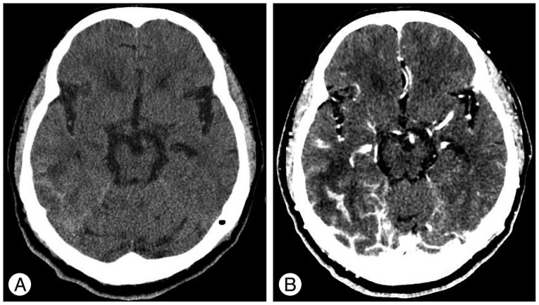
Fig. 1 Brain CT shows diffuse high density lesion in the base of right temporal lobe which is similar finding with subarachnoid hemorrhage on non-contrast image (A). Strong enhancements with multiple, branching, linear pattern are seen in the same area (B).
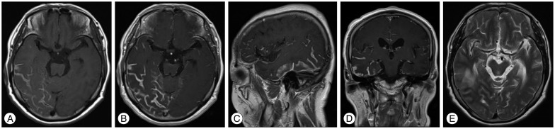
Fig. 2 Brain MRI shows multiple, branching, linear high signal intensity lesions in the right temporal base on axial T1-WI (A), and also strong enhancements with same pattern are seen (B). These findings are mainly found in the lateral and inferior surfaces of the temporal lobe on enhanced sagittal (C) and coronal T1-WIs (D). Low signal intensity lesions with same patterns are seen in the same location on axial T2-WI (E).
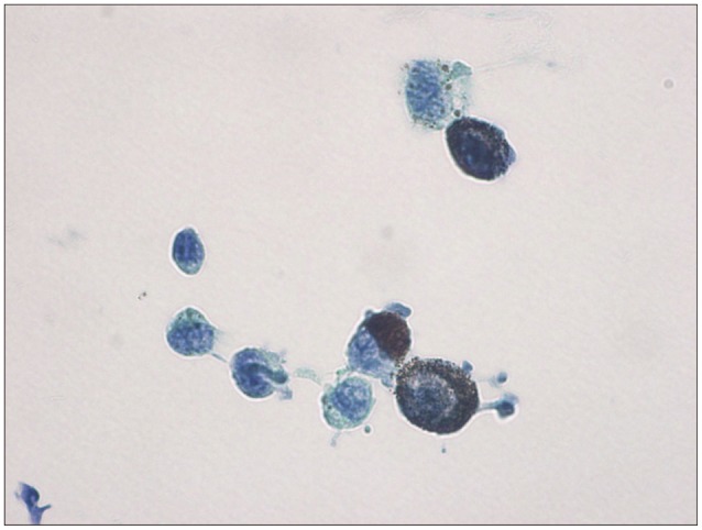
Fig. 3 Pathological findings shows pleomorphic cells with abundant cytoplasm, abundant intracytoplasmic melanin pigments, nuclear pleomorphism, and prominent nucleoli (Papanicolaou stain, ×1000). These tumor cells showed strong immunoreactivity to HMB-45, S-100, and Melan-A on the immunohistochemical study.
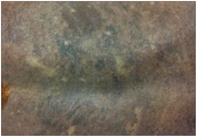
Fig. 4 Photograph of skin lesion on the back. This lesion was confirmed as pigmented nevi through the biopsy.
Reference
-
1. Gempt J, Buchmann N, Grams AE, Zoubaa S, Schlegel J, Meyer B, et al. Black brain : transformation of a melanocytoma with diffuse melanocytosis into a primary cerebral melanoma. J Neurooncol. 2011; 102:323–328. PMID: 20640479.
Article2. Greco Crasto S, Soffietti R, Bradac GB, Boldorini R. Primitive cerebral melanoma : case report and review of the literature. Surg Neurol. 2001; 55:163–168. discussion 168PMID: 11311915.3. Hayward RD. Malignant melanoma and the central nervous system. A guide for classification based on the clinical findings. J Neurol Neurosurg Psychiatry. 1976; 39:526–530. PMID: 950562.
Article4. Ibáñez J, Weil B, Ayala A, Jimenez A, Acedo C, Rodrigo I. Meningeal melanocytoma : case report and review of the literature. Histopathology. 1997; 30:576–581. PMID: 9205863.5. Jaiswal S, Vij M, Tungria A, Jaiswal AK, Srivastava AK, Behari S. Primary melanocytic tumors of the central nervous system : a neuroradiological and clinicopathological study of five cases and brief review of literature. Neurol India. 2011; 59:413–419. PMID: 21743173.
Article6. Jellinger K, Chou P, Paulus W. Melanocytic lesions. In : Kleihues P, Cavanee WK, editors. Pathology and Genetics of Tumours of the Nervous system. Lyon: IARC Press;2000.7. Lee CJ, Rhee DY, Heo W, Park HS. Primary leptomeningeal malignant melanoma. J Korean Neurosurg Soc. 2004; 36:425–427.8. Louis DN, Ohgaki H, Wiestler OD, Cavenee WK, Burger PC, Jouvet A, et al. The 2007 WHO classification of tumours of the central nervous system. Acta Neuropathol. 2007; 114:97–109. PMID: 17618441.
Article9. Piedra MP, Scheithauer BW, Driscoll CL, Link MJ. Primary melanocytic tumor of the cerebellopontine angle mimicking a vestibular schwannoma : case report. Neurosurgery. 2006; 59:E206. discussion E206. PMID: 16823290.10. Pirini MG, Mascalchi M, Salvi F, Tassinari CA, Zanella L, Bacchini P, et al. Primary diffuse meningeal melanomatosis : radiologic-pathologic correlation. AJNR Am J Neuroradiol. 2003; 24:115–118. PMID: 12533338.11. Roser F, Nakamura M, Brandis A, Hans V, Vorkapic P, Samii M. Transition from meningeal melanocytoma to primary cerebral melanoma. Case report. J Neurosurg. 2004; 101:528–531. PMID: 15352613.12. Shah I, Imran M, Akram R, Rafat S, Zia K, Emaduddin M. Primary intracranial malignant melanoma. J Coll Physicians Surg Pak. 2013; 23:157–159. PMID: 23374525.
- Full Text Links
-
- Actions
-
Cited
- CITED
-
- Close
- Share
-
- Similar articles
-
- A Case of Cerebral Leptomeningeal Melanomatosis Associated with Large Hairly Nevi in Adult
- A Case of Primary Malignant Leptomeningeal Melanomatosis
- Primary Leptomeningeal Malignant Melanoma
- A Case of Leptomeningeal Metastasis Associated with Cerebral Venous Thrombosis
- Malignant Ascites after Subduroperitoneal Shunt in a Patient with Leptomeningeal Metastasis
