Ann Surg Treat Res.
2016 Jan;90(1):16-20. 10.4174/astr.2016.90.1.16.
Stereotactic vacuum-assisted breast biopsy under lateral decubitus position
- Affiliations
-
- 1Department of Breast and Thyroid Surgery, Research Institute for Convergence of Biomedical Science and Technology, Pusan National University Yangsan Hospital, Yangsan, Korea. isepa102@naver.com
- 2Department of Pathology, Pusan National University Yangsan Hospital, Yangsan, Korea.
- 3Department of Radiology, Pusan National University Yangsan Hospital, Yangsan, Korea.
- KMID: 2149927
- DOI: http://doi.org/10.4174/astr.2016.90.1.16
Abstract
- PURPOSE
Stereotactic vacuum-assisted breast biopsy (VAB) has been established as a standard method for histological diagnosis of microcalcification or nonpalpable breast lesions on mammography. Generally, the procedure has been done under the prone position or upright sitting position. We herein attempt to evaluate clinical utility of Stereotactic VAB under lateral decubitus position.
METHODS
One hundred six women (mean age, 51.2 years) with mammographically detected microcalcification underwent lateral decubitus positioning VAB using the 8G probe. In all cases, we obtained mammography specimens for identification of microcalcification and postprocedure mammography. We reviewed mean procedure time, pieces of specimen, pathology and follow-up mammography.
RESULTS
The procedure took approximately 20 minutes (range, 15-24 minutes). Average number of obtained specimens was 8.5 pieces (range, 6-12 pieces). Microcalcifications were confirmed in both specimen mammography and microscopic slides. Of 106 cases, 10 cases were diagnosed as ductal carcinoma in situ. Additional surgical management was performed. Atypical ductal hyperplasias were found in 8 cases, and fibrocystic changes in 88 cases.
CONCLUSION
Stereotactic VAB using the 8G probe under lateral decubitus position does not need a dedicated table, and is easier to maintain the position. Also, this procedure is accurate and safe. Thus, stereotactic VAB using the 8G probe under lateral decubitus position will be a useful method for diagnosis of microcalcification or nonpalpable breast lesions on mammography.
MeSH Terms
Figure
-

Fig. 1 (A) Patient laid on the stereotactic vacuum-assisted breast biopsy table in the lateral decubitus position. (B) The breast is compressed and fixed to the appropriate direction.
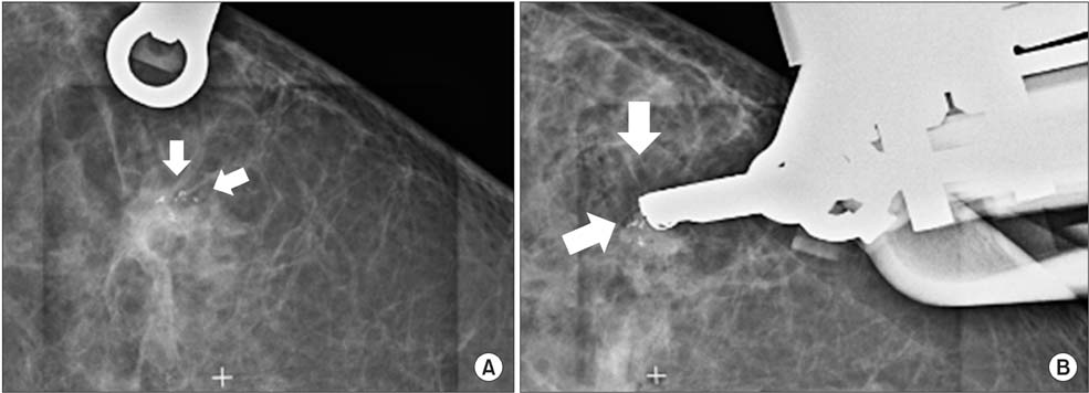
Fig. 2 (A) Mammography for confirmation of exact location of needle after introduction (arrows: microcalcification). (B) Mammography for confirmation of exact location of needle after introduction (arrows: microcalcification).

Fig. 3 Specimen mammography shows a microcalcification (arrow) two separate specimens.
Reference
-
1. Korean Central Cancer Registry. 2002 Annual report of the Korean Central Cancer Registry. Gwacheon: Ministry of Health and Welfare;2003.2. Bae JW, Koo BW. Retrospective analysis of the needle localization biopsies for nonpalpable mammographic microcalcifications of the breast. J Korean Surg Soc. 1994; 46:335–341.3. Kerlikowske K, Grady D, Rubin SM, Sandrock C, Ernster VL. Efficacy of screening mammography. A meta-analysis. JAMA. 1995; 273:149–154.4. Reynolds HE, Poon CM, Goulet RJ, Lazaridis CL. Biopsy of breast microcalcifications using an 11-gauge directional vacuum-assisted device. AJR Am J Roentgenol. 1998; 171:611–613.5. Meyer JE, Smith DN, DiPiro PJ, Denison CM, Frenna TH, Harvey SC, et al. Stereotactic breast biopsy of clustered microcalcifications with a directional, vacuum-assisted device. Radiology. 1997; 204:575–576.6. Burak WE Jr, Owens KE, Tighe MB, Kemp L, Dinges SA, Hitchcock CL, et al. Vacuumassisted stereotactic breast biopsy: histologic underestimation of malignant lesions. Arch Surg. 2000; 135:700–703.7. Rotter K, Haentschel G, Koethe D, Goetz L, Bornhofen-Poschke A, Lebrecht A, et al. Evaluation of mammographic and clinical follow-up after 755 stereotactic vacuum-assisted breast biopsies. Am J Surg. 2003; 186:134–142.8. Walker TM. Impalpalbe breast lesions: stereotactic core biopsy with 'add-on' unit. The Breast. 1997; 6:126–131.9. Kirshenbaum KJ, Voruganti T, Overbeeke C, Kirshenbaum MD, Patel P, Kaplan G, et al. Stereotactic core needle biopsy of nonpalpable breast lesions using a conventional mammography unit with an add-on device. AJR Am J Roentgenol. 2003; 181:527–531.10. Welle GJ, Clark M, Loos S, Pauls D, Warden D, Sheffield M, et al. Stereotactic breast biopsy: recumbent biopsy using add-on upright equipment. AJR Am J Roentgenol. 2000; 175:59–63.11. Burbank F. Stereotactic breast biopsy: comparison of 14- and 11-gauge Mammotome probe performance and complication rates. Am Surg. 1997; 63:988–995.12. Philpotts LE, Lee CH, Horvath LJ, Tocino I. Canceled stereotactic core-needle biopsy of the breast: analysis of 89 cases. Radiology. 1997; 205:423–428.
- Full Text Links
-
- Actions
-
Cited
- CITED
-
- Close
- Share
-
- Similar articles
-
- Lateral Decubitus Positioning Stereotactic Vacuum-Assisted Breast Biopsy with True Lateral Mammography
- The Efficacy of Stereotactic Vacuum-assisted Biopsy and Needle Localization Vacuum-assisted Biopsy for Diagnosing Breast Microcalcification
- The Clinical Utility of a Adding Lateral Approach to Conventional Vertical Approach for Prone Stereotactic Vacuum-Assisted Breast Biopsy
- Diagnosis of Non-palpable Breast Lesions with Microcalcification by Upright Add-on Type Stereotactic Vacuum-assisted Biopsy
- Feasibility of Stereotactic Biopsy for Breast Lesions with the Patient in the Decubitus Position: Our Early Experience
