The Clinical Utility of a Adding Lateral Approach to Conventional Vertical Approach for Prone Stereotactic Vacuum-Assisted Breast Biopsy
- Affiliations
-
- 1Department of Radiology, Seoul St. Mary's Hospital, College of Medicine, The Catholic University of Korea, Seoul 137-040, Korea. lionmain@catholic.ac.kr
- KMID: 1715760
- DOI: http://doi.org/10.3348/kjr.2013.14.4.568
Abstract
OBJECTIVE
The purpose of this study is to evaluate the clinical utility of adding lateral approach to conventional vertical approach for prone stereotactic vacuum-assisted breast biopsies.
MATERIALS AND METHODS
From April 2010 to May 2012, 130 vacuum-assisted stereotactic biopsies were attempted in 127 patients. While a vertical approach was preferred, a lateral approach was used if the vertical approach failed. The success rate of biopsies utilizing only a vertical approach was compared with that using both vertical and lateral approaches and the breast thickness for both procedures was measured and compared with that for vertical approach. In addition, pathology results were evaluated and the causes of the failed biopsies were analyzed.
RESULTS
Of the 130 cases, 127 biopsies were performed and 3 biopsies failed. The success rate of the vertical approach was 83.8% (109/130); however, when the lateral approach was also used, the success rate increased to 97.7% (127/130) (p = 0.0004). The mean breast thickness was 2.7 +/- 1 cm for the lateral approach and 4 +/- 1.2 cm for the vertical approach (p < 0.0001). The histopathologic results in 76 (59.8%) of the biopsies were benign, 23 (18.1%) were high-risk lesions, and 28 (22.0%) were malignant. The causes of biopsy failure were thin breasts (n = 2) and undetected difficult lesion location (n = 1).
CONCLUSION
The addition of lateral approach to conventional vertical approach in prone stereotactic vacuum-assisted breast biopsy improved the success rate of stereotactic biopsy, especially in patients with thin breasts.
MeSH Terms
Figure
-
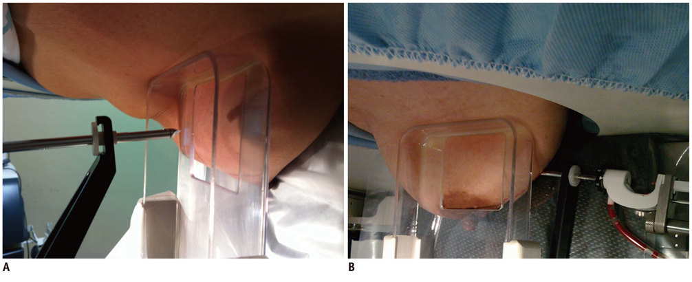
Fig. 1 Vertical and lateral approaches. A. Vertical approach for stereotactic biopsy using prone-type device. B. Lateral approach for stereotactic biopsy using prone-type device.

Fig. 2 Fibrocystic change in 41-year-old woman was confirmed by lateral approach. Mediolateral oblique view of mammography (A) and spot view before lateral approach of stereotactic biopsy (B) showing clustered, amorphous and punctate microcalcifications in left breast (arrows). Breast thickness was measured as 1.9 cm at this time. Spot view after inserting needle (C), spot view after biopsy (D), and spot view of specimen (E) were seen. Because there were several microcalcifications in more than 3 specimens (arrows), this procedure was defined as success, and fibrocystic change was confirmed.
Reference
-
1. Berg WA, Krebs TL, Campassi C, Magder LS, Sun CC. Evaluation of 14- and 11-gauge directional, vacuum-assisted biopsy probes and 14-gauge biopsy guns in a breast parenchymal model. Radiology. 1997; 205:203–208.2. Elvecrog EL, Lechner MC, Nelson MT. Nonpalpable breast lesions: correlation of stereotaxic large-core needle biopsy and surgical biopsy results. Radiology. 1993; 188:453–455.3. Parker SH, Lovin JD, Jobe WE, Burke BJ, Hopper KD, Yakes WF. Nonpalpable breast lesions: stereotactic automated largecore biopsies. Radiology. 1991; 180:403–407.4. Reynolds HE, Poon CM, Goulet RJ, Lazaridis CL. Biopsy of breast microcalcifications using an 11-gauge directional vacuum-assisted device. AJR Am J Roentgenol. 1998; 171:611–613.5. Nakamura Y, Urashima M, Matsuura A, Nishihara R, Itoh A, Kagemoto M, et al. Stereotactic directional vacuum-assisted breast biopsy using lateral approach. Breast Cancer. 2010; 17:286–289.6. Han BK, Choe YH, Ko YH, Nam SJ, Kim JH, Yang JH. Stereotactic core-needle biopsy of non-mass calcifications: outcome and accuracy at long-term follow-up. Korean J Radiol. 2003; 4:217–223.7. Lehman CD, Sieler-Gutierrez HJ, Georgian-Smith D. Lateral approach biopsy adapter: accuracy on an upright unit in a turkey breast model. AJR Am J Roentgenol. 2001; 177:897–899.8. American College of Radiology. Breast imaging reporting and data system (BI-RADS). 4th ed. Reston, VA: American College of Radiology;2004.9. Stomper PC, Connolly JL, Meyer JE, Harris JR. Clinically occult ductal carcinoma in situ detected with mammography: analysis of 100 cases with radiologic-pathologic correlation. Radiology. 1989; 172:235–241.10. Spencer NJ, Evans AJ, Galea M, Sibbering DM, Yeoman LJ, Pinder SE, et al. Pathological-radiological correlations in benign lesions excised during a breast screening programme. Clin Radiol. 1994; 49:853–856.11. Soo MS, Baker JA, Rosen EL. Sonographic detection and sonographically guided biopsy of breast microcalcifications. AJR Am J Roentgenol. 2003; 180:941–948.12. Jackman RJ, Marzoni FA Jr. Stereotactic histologic biopsy with patients prone: technical feasibility in 98% of mammographically detected lesions. AJR Am J Roentgenol. 2003; 180:785–794.13. Philpotts LE, Shaheen NA, Carter D, Lange RC, Lee CH. Comparison of rebiopsy rates after stereotactic core needle biopsy of the breast with 11-gauge vacuum suction probe versus 14-gauge needle and automatic gun. AJR Am J Roentgenol. 1999; 172:683–687.14. Jackman RJ, Burbank F, Parker SH, Evans WP 3rd, Lechner MC, Richardson TR, et al. Stereotactic breast biopsy of nonpalpable lesions: determinants of ductal carcinoma in situ underestimation rates. Radiology. 2001; 218:497–502.15. Meyer JE, Smith DN, Lester SC, Kaelin C, DiPiro PJ, Denison CM, et al. Large-core needle biopsy of nonpalpable breast lesions. JAMA. 1999; 281:1638–1641.16. Kim HS, Kim MJ, Kim EK, Kwak JY, Son EJ, Oh KK. US-guided vacuum-assisted biopsy of microcalcifications in breast lesions and long-term follow-up results. Korean J Radiol. 2008; 9:503–509.17. Welle GJ, Clark M, Loos S, Pauls D, Warden D, Sheffield M, et al. Stereotactic breast biopsy: recumbent biopsy using add-on upright equipment. AJR Am J Roentgenol. 2000; 175:59–63.18. Kettritz U, Morack G, Decker T. Stereotactic vacuum-assisted breast biopsies in 500 women with microcalcifications: radiological and pathological correlations. Eur J Radiol. 2005; 55:270–276.19. Youk JH, Kim EK, Kim MJ, Ko KH, Kwak JY, Son EJ, et al. Concordant or discordant? Imaging-pathology correlation in a sonography-guided core needle biopsy of a breast lesion. Korean J Radiol. 2011; 12:232–240.
- Full Text Links
-
- Actions
-
Cited
- CITED
-
- Close
- Share
-
- Similar articles
-
- Lateral Decubitus Positioning Stereotactic Vacuum-Assisted Breast Biopsy with True Lateral Mammography
- The Efficacy of Stereotactic Vacuum-assisted Biopsy and Needle Localization Vacuum-assisted Biopsy for Diagnosing Breast Microcalcification
- Stereotactic vacuum-assisted breast biopsy under lateral decubitus position
- Diagnosis of Non-palpable Breast Lesions with Microcalcification by Upright Add-on Type Stereotactic Vacuum-assisted Biopsy
- Treating Gynecomastia with Ultrasound-guided Vacuum-assisted Biopsy Device as a Cosmetic Method
