J Korean Soc Radiol.
2011 Jan;64(1):75-81. 10.3348/jksr.2011.64.1.75.
Feasibility of Stereotactic Biopsy for Breast Lesions with the Patient in the Decubitus Position: Our Early Experience
- Affiliations
-
- 1Department of Radiology, Yonsei University College of Medicine, Korea. mines@yuhs.ac
- KMID: 1443586
- DOI: http://doi.org/10.3348/jksr.2011.64.1.75
Abstract
- PURPOSE
We performed add-on type stereotactic vaccum-assisted breast biopsy (SVABB) with the patient in the decubitus position and we determined the feasibility of SVABB according to the breast thickness, the vicinity of the lesion in relation to the chest wall and the histopathologic diagnosis, and we report here on our initial experience.
MATERIALS AND METHODS
81 patients with 82 lesions underwent SVABB. We measured the distance of the lesion from the chest wall on the basis of mammography, and we evaluated the breast thickness on the basis of the magnified, stereotactic view. The histopathologic results and the rate of underestimating atypical findings and ductal carcinoma in situ were analyzed.
RESULTS
The range of breast thickness was 12~61 mm on the magnified view and the range of breast thickness was 16~55 mm on the stereotactic view. The range of the distance of the lesion to the chest wall was 0~67 mm. The histopathologic results of SVABB demonstrated benign findings for 62 lesions, atypical findings for 10 lesions, ductal carcinoma in situ for 8 lesions and invasive ductal carcinoma for 2 lesions. The estimated underestimation rate was 30% (3/10) for the atypical finding and 0% (0/7) for ductal carcinoma in situ.
CONCLUSION
This study demonstrated the results of add-on type SVABB with the patient in the decubitus position and SVABB was successful for patients with various lesion locations and breast thicknesses.
MeSH Terms
Figure
-
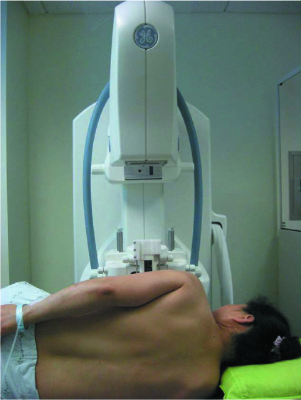
Fig. 1 Stereotatic vaccum assisted biopsy was performed on decubitus position.
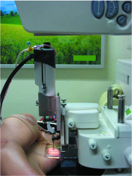
Fig. 2 Directional vaccum-assisted biopsy probe inserted perpendicular direction in relation to the compressed breast tissue plane.
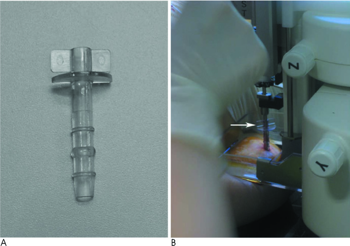
Fig. 3 To prevent of skin tear during the procedure, plastic skin protector (A, arrow in B) was applied.
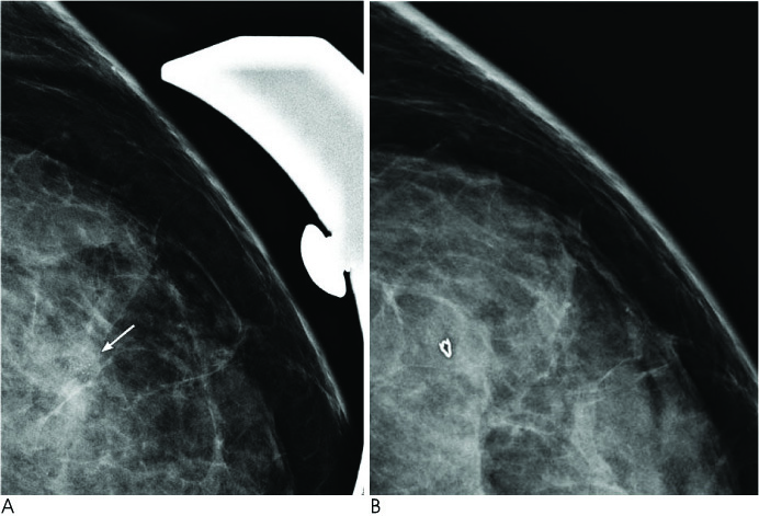
Fig. 4 A. Suspicious clustered microcalcifications(arrow) were depicted on magnification mammography. B. Follow up magnification mammographic view after stereotactic biopy, marker inserted at calcification retrieval site.
Reference
-
1. Parker SH, Lovin JD, Jobe WE, Luethke JM, Hopper KD, Yakes WF, et al. Stereotactic breast biopsy with a biopsy gun. Radiology. 1990; 176:741–747.2. Lee CH, Egglin TK, Philpotts L, Mainiero MB, Tocino I. Cost-effectiveness of stereotactic core needle biopsy: analysis by means of mammographic findings. Radiology. 1997; 202:849–854.3. Brenner RJ, Bassett LW, Fajardo LL, Dershaw DD, Evans WP 3rd, Hunt R, et al. Stereotactic core-needle breast biopsy: a multi-institutional prospective trial. Radiology. 2001; 218:866–872.4. Liberman L, Sama MP. Cost-effectiveness of stereotactic 11-gauge directional vacuum-assisted breast biopsy. AJR Am J Roentgenol. 2000; 175:53–58.5. Kirshenbaum KJ, Voruganti T, Overbeeke C, Kirshenbaum MD, Patel P, Kaplan G, et al. Stereotactic core needle biopsy of nonpalpable breast lesions using a conventional mammography unit with an add-on device. AJR Am J Roentgenol. 2003; 181:527–531.6. Han BK, Choe YH, Ko YH, Nam SJ, Kim JH, Yang JH. Stereotactic core-needle biopsy of non-mass calcifications: outcome and accuracy at long-term follow-up. Korean J Radiol. 2003; 4:217–223.7. Seo MR, Park JM, Gong GY, Ahn SH. Results with Add-on Stereotactic Core Biopsy (ASCB) of the Breast Lesions. J Korean Radiol Soc. 2000; 43:245–250.8. American College of Radiology. Breast imaging reporting and data system (BI-RADS). 4th ed. Reston. VA.: America College of Radiology;2003.9. Liberman L, Feng TL, Dershaw DD, Morris EA, Abramson AF. US-guided core breast biopsy: use and cost-effectiveness. Radiology. 1998; 208:717–723.10. Dillon MF, Hill AD, Quinn CM, O'Doherty A, McDermott EW, O'Higgins N. The accuracy of ultrasound, stereotactic, and clinical core biopsies in the diagnosis of breast cancer, with an analysis of false-negative cases. Ann Surg. 2005; 242:701–707.11. Fajardo LL, Pisano ED, Caudry DJ, Gatsonis CA, Berg WA, Connolly J, et al. Stereotactic and sonographic large-core biopsy of nonpalpable breast lesions: results of the Radiologic Diagnostic Oncology Group V study. Acad Radiol. 2004; 11:293–308.12. Caines JS, McPhee MD, Konok GP, Wright BA. Stereotaxic needle core biopsy of breast lesions using a regular mammographic table with an adaptable stereotaxic device. AJR Am J Roentgenol. 1994; 163:317–321.13. Levin MF, Papoff WJ, Doan L, Eliasziw M. Stereotaxic percutaneous core biopsy versus surgical biopsy of nonpalpable breast lesions using a standard mammographic table with an add-on device. Can Assoc Radiol J. 2001; 52:29–32.14. Becker L, Taves D, McCurdy L, Muscedere G, Karlik S, Ward S. Stereotactic core biopsy of breast microcalcifications: comparison of film versus digital mammography, both using an add-on unit. AJR Am J Roentgenol. 2001; 177:1451–1457.15. Nakamura Y, Urashima M, Matsuura A, Nishihara R, Itoh A, Kagemoto M, et al. Stereotactic directional vacuum-assisted breast biopsy using lateral approach. Breast Cancer. 2010; 17:286–289.16. Liberman L, Smolkin JH, Dershaw DD, Morris EA, Abramson AF, Rosen PP. Calcification retrieval at stereotactic, 11-gauge, directional, vacuum-assisted breast biopsy. Radiology. 1998; 208:251–260.17. Kim SH, Lee JH, Song BJ, Jung SS. Stereotactic vacuum-assisted biopsy of microcalcifications using an upright add-on type stereotactic mammography unit. J Korean Radiol Soc. 2007; 57:291–297.18. Liberman L, Evans WP 3rd, Dershaw DD, Hann LE, Deutch BM, Abramson AF, et al. Radiography of microcalcifications in stereotaxic mammary core biopsy specimens. Radiology. 1994; 190:223–225.19. Philpotts LE, Shaheen NA, Carter D, Lange RC, Lee CH. Comparison of rebiopsy rates after stereotactic core needle biopsy of the breast with 11-gauge vacuum suction probe versus 14-gauge needle and automatic gun. AJR Am J Roentgenol. 1999; 172:683–687.20. Sohn V, Arthurs Z, Herbert G, Keylock J, Perry J, Eckert M, et al. Atypical ductal hyperplasia: improved accuracy with the 11-gauge vacuum-assisted versus the 14-gauge core biopsy needle. Ann Surg Oncol. 2007; 14:2497–2501.21. Pfarl G, Helbich TH, Riedl CC, Wagner T, Gnant M, Rudas M, et al. Stereotactic 11-gauge vacuum-assisted breast biopsy: a validation study. AJR Am J Roentgenol. 2002; 179:1503–1507.22. Won B, Reynolds HE, Lazaridis CL, Jackson VP. Stereotactic biopsy of ductal carcinoma in situ of the breast using an 11-gauge vacuum-assisted device: persistent underestimation of disease. AJR Am J Roentgenol. 1999; 173:227–229.23. Bagnall MJ, Evans AJ, Wilson AR, Burrell H, Pinder SE, Ellis IO. When have mammographic calcifications been adequately sampled at needle core biopsy? Clin Radiol. 2000; 55:548–553.24. Kettritz U, Rotter K, Schreer I, Murauer M, Schulz-Wendtland R, Peter D, et al. Stereotactic vacuum-assisted breast biopsy in 2874 patients: a multicenter study. Cancer. 2004; 100:245–251.
- Full Text Links
-
- Actions
-
Cited
- CITED
-
- Close
- Share
-
- Similar articles
-
- Lateral Decubitus Positioning Stereotactic Vacuum-Assisted Breast Biopsy with True Lateral Mammography
- Stereotactic vacuum-assisted breast biopsy under lateral decubitus position
- Mammography-Guided Interventional Procedure
- The Utility of ABBI (Advanced breast biopsy instrumentation) for Non-palpable Breast Lesions
- The Efficacy of Stereotactic Vacuum-assisted Biopsy and Needle Localization Vacuum-assisted Biopsy for Diagnosing Breast Microcalcification
