Tracking of Neural Stem Cells in Rats with Intracerebral Hemorrhage by the Use of 3T MRI
- Affiliations
-
- 1Department of Radiology, Chonnam National University Medical School, Gwang-ju, Korea. yjeong@jnu.ac.kr
- 2Department of Physiology, Chonnam National University Medical School, Gwang-ju, Korea.
- 3Department of Pathology, Chonnam National University Medical School, Gwang-ju, Korea.
- KMID: 1758452
- DOI: http://doi.org/10.3348/kjr.2008.9.3.196
Abstract
OBJECTIVE
To access the feasibility of clinically available 3T MRI to detect the migration of labeled neural stem cells (NSCs) in intracerebral hemorrhage (ICH) in a rat model. MATERIALS AND METHODS: The ethics committee of our institution approved this study. ICH was induced by the injection of collagenase type IV into the right striatum of ten Sprague-Dawley rats. Human NSCs conjugated with Feridex (super-paramagnetic iron oxide: SPIO) were transplanted into the left striatum one week after ICH induction. MRI was performed on a 3T scanner during the first, second, third, fourth, and sixth weeks post-transplantation. MRI was obtained using coronal T2- and T2*-weighted sequences. Two rats were sacrificed every week after in vivo MRI in order to analyze the histological findings. RESULTS: ICH in the right striatum was detected by MRI one and two weeks after transplantation without migration of the NSCs. There was no migration of the NSCs as seen on the histological findings one week after transplantation. The histological findings two weeks after transplantation showed a small number of NSCs along the corpus callosum. On MRI three weeks after transplantation, there was a hypointense line along the corpus callosum and decreased signal intensity in the right periventricular region. Histological findings three weeks after transplantation confirmed the presence of the hypointense line representing SPIO-labeled NSCs. MRI four and six weeks after transplantation showed a hypointense spot in the right periventricular region. The histological findings four and six weeks after transplantation showed the presence of prominent NSCs in the right periventricular region. CONCLUSION: 3T MRI can detect the migration of NSCs in rats with ICH along the corpus callosum. Therefore, 3T MRI could be feasible for detecting the migration of NSCs in the clinical setting of stem cell therapy.
Keyword
MeSH Terms
Figure
-
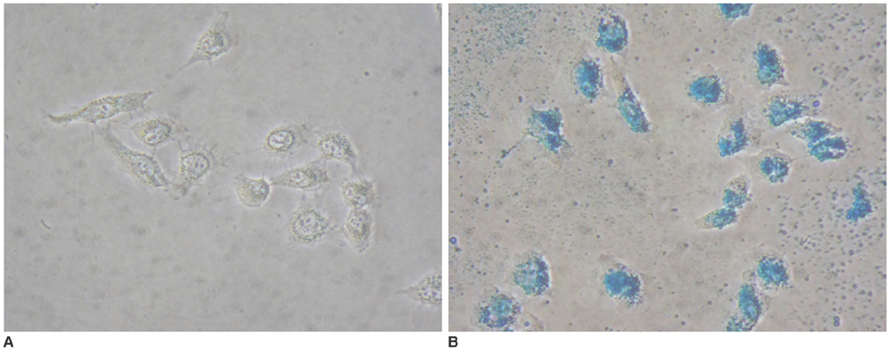
Fig. 1 Prussian blue staining of neural stem cells (objective magnification: × 40). A. Unlabeled neural stem cells without blue spots. B. SPIO-labeled neural stem cells show blue spots located inside cells, suggesting presence of iron oxide particles.
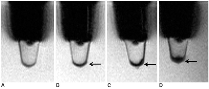
Fig. 2 In vitro T2*-weighted images of neural stem cells. A-D. 250 neural stem cells in 2% gelatin tube (A), 5 × 10³ neural stem cells (B), 5 × 10⁴ neural stem cells (C), 2 × 10⁵ neural stem cells (D). In B, C, and D, T2*-weighted images (TR/TE = 231/10 msec) show linear, hypointense cluster in bottom of tubes (arrows), representing SPIO-labeled neural stem cells.
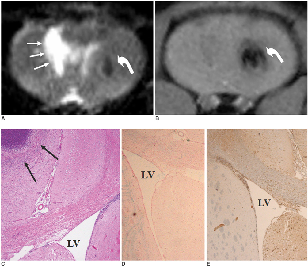
Fig. 3 MR and histological findings one week after implantation of neural stem cells. A. T2-weighted image (TR/TE = 3,000/100 msec) shows hypointense, SPIO-labeled, neural stem cells (curved arrow) in left striatum and hyperintense lesion (arrows) in right striatum, representing intracerebral hemorrhage. B. T2*-weighted image (TR/TE = 231/10 msec) reveals hypointense, SPIO-labeled neural stem cells (curved arrow) in left cerebral hemisphere. C. Hematoxylin and eosin staining (objective magnification: × 4) shows intracerebral hemorrhage (arrows) in right striatum with mass effect to corpus callosum and right lateral ventricle (LV). D. Prussian blue staining (objective magnification: × 4) shows no migration of SPIO-labeled neural stem cells. E. Immunohistochemical staining (objective magnification: × 4) shows no migration of neural stem cells.
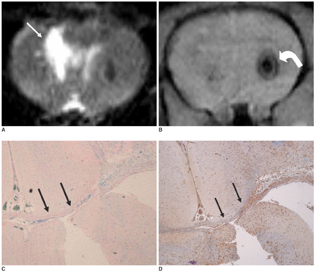
Fig. 4 MR and histological findings two weeks after implantation of neural stem cells. A. On T2-weighted image (TR/TE = 3,000/100 msec), size of intracerebral hemorrhage (arrow) in right cerebral hemisphere is smaller than it was one week after implantation of neural stem cells. B. T2*-weighted image (TR/TE = 231/10 msec) shows slightly decreased size of hypointense spot (curved arrow) in left cerebral hemisphere. There is no migration of neural stem cells. C. Prussian blue staining (objective magnification: × 4) shows small number of neural stem cells in corpus callosum (arrows). D. Immunohistochemical staining (objective magnification: × 4) shows migration of small number of neural stem cells into corpus callosum (arrows).
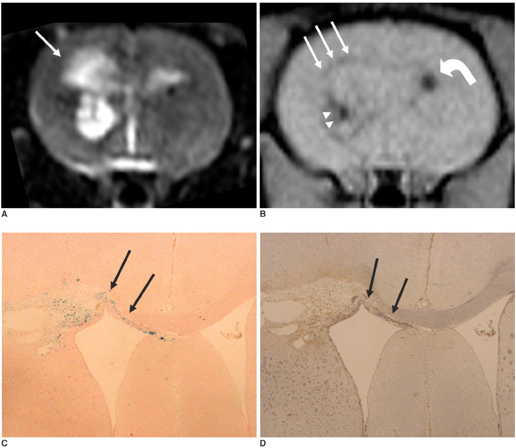
Fig. 5 MR and histological findings three weeks after implantation of neural stem cells. A. T2-weighted image (TR/TE = 3,000/100 msec) shows decreased size (arrow) of intracerebral hemorrhage two weeks after implantation of neural stem cells. B. T2*-weighted image (TR/TE = 231/10 msec) shows decreased size of hypointense spot (curved arrow) in left striatum. Linear low signal intensity (arrows) along corpus callosum is considered to represent SPIO-labeled neural stem cells. There is small, hypointense spot in right, periventricular region (arrowheads), suggestive of migrated neural stem cells. C. Prussian blue staining (objective magnification: × 2) shows large number of SPIO-labeled, neural stem cells in corpus callosum (arrows). D. Immunohistochemical staining (objective magnification: × 2) shows neural stem cells in corpus callosum (arrows).
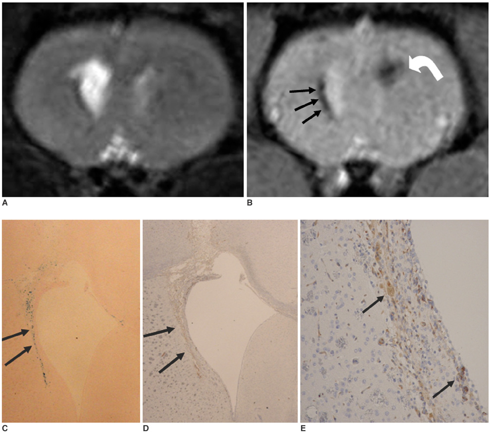
Fig. 6 MR and histological findings four weeks after implantation of neural stem cells. A. T2-weighted image (TR/TE = 3,000/100 msec) shows decreased size of intracerebral hemorrhage three weeks after implantation of neural stem cells. B. T2*-weighted image (TR/TE = 231/10 msec) shows decreased size of hypointense spot (curved arrow) in left striatum. Clusters of hypointensity (arrows) are seen in right periventricular region. C. Prussian blue staining (objective magnification: × 4) shows inhomogeneous distribution of neural stem cells around right ventricle (arrows). D, E. Immunohistochemical staining (objective magnification: × 4 and × 10) shows neural stem cells in right, periventricular region (arrows).
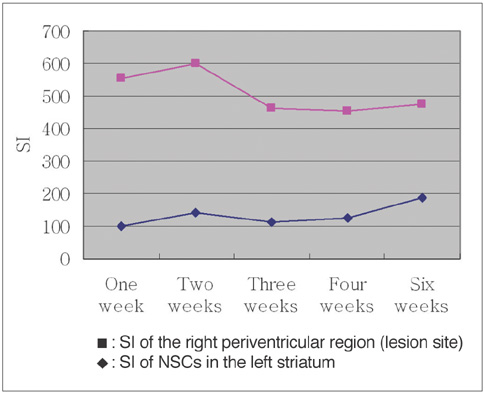
Fig. 7 Signal intensity ratio changes in MR images of right periventricular region and of left striatum. Significant decrease of signal intensity ratio between two weeks and three weeks after implantation of neural stem cells is seen in right periventricular region, suggesting massive migration of SPIO-labeled neural stem cells from left striatum into right periventricular region.
Cited by 1 articles
-
A Study of Feasibility of Brain Imaging in Medium- and Small-Sized Animals: Using a Clinical 3T MR System with Three Surface Coils
Shin Young Park, Mi Ri Jeong, Byung Mann Cho, Kang Soo Kim, Hak Jin Kim
J Korean Soc Radiol. 2017;77(5):317-326. doi: 10.3348/jksr.2017.77.5.317.
Reference
-
1. Aboody KS, Brown A, Rainov NG, Bower KA, Liu S, Yang W, et al. Neural stem cells display extensive tropism for pathology in adult brain: evidence from intracranial gliomas. Proc Natl Acad Sci USA. 2000. 97:12846–12851.2. Modo M, Stroemer RP, Tang E, Patel S, Hodges H. Effects of implantation site of stem cell grafts on behavioral recovery from stroke damage. Stroke. 2002. 33:2270–2278.3. Bjorklund A, Lindvall O. Cell replacement therapies for central nervous system disorders. Nat Neurosci. 2000. 3:537–544.4. Savitz SI, Rosenbaum DM, Dinsmore JH, Wechsler LR, Caplan LR. Cell transplantation for stroke. Ann Neurol. 2002. 52:266–275.5. Qureshi AI, Tuhrim S, Broderick JP, Batjer HH, Hondo H, Hanley DF. Spontaneous intracerebral hemorrhage. N Engl J Med. 2001. 344:1450–1460.6. Lo YK, Yiu Ch, Hu HH, Su MS, Laeuchli SC. Frequency and characteristics of early seizures in Chinese acute stroke. Acta Neurol Scand. 1994. 90:83–85.7. Jeong SW, Chu K, Jung KH, Kim SU, Kim M, Roh JK. Human neural stem cell transplantation promotes functional recovery in rats with experimental intracerebral hemorrhage. Stroke. 2003. 34:2258–2263.8. Kim DE, Schellingerhout D, Ishii K, Shah K, Weissleder R. Imaging of stem cell recruitment to ischemic infarcts in a murine model. Stroke. 2004. 35:952–957.9. Hoehn M, Kustermann E, Blunk J, Wiedermann D, Trapp T, Wecker S, et al. Monitoring of implanted stem cell migration in vivo: a highly resolved in vivo magnetic resonance imaging investigation of experimental sroke in rat. Proc Natl Acad Sci USA. 2002. 99:16267–16272.10. Magnitsky S, Watson DJ, Walton RM, Pickup S, Bulte JW, Wolfe JH, et al. In vivo and ex vivo MRI detection of localized and disseminated neural stem cell grafts in the mouse brain. Neuroimage. 2005. 26:744–754.11. Modo M, Mellodew K, Cash D, Fraser SE, Meade TJ, Price J, et al. Mapping transplanted stem cell migration after a stroke: a serial, in vivo magnetic resonance imaging study. Neuroimage. 2004. 21:311–317.12. Bang OY, Lee JS, Lee PH, Lee G. Autologous mesenchymal stem cell transplantation in stroke patients. Ann Neurol. 2005. 57:874–882.13. Zhang Z, Ziang Q, Jaing F, Ding G, Zhang R, Wang L, et al. In vivo magnetic resonance imaging tracks adult neural progenitor cell targeting of brain tumor. Neuroimage. 2004. 23:281–287.14. Frank JA, Miller BR, Arbab AS, Zuwicke H, Jordan EK, Lewis BK, et al. Clinically applicable labeling of mammalian and stem cells by combining superparamagnetic iron oxides and transfection agents. Radiology. 2003. 228:480–487.15. Bulte JW, Duncan ID, Frank JA. In vivo magnetic resonance tracking of magnetically labeled cells after transplantation. J Cereb Blood Flow Metab. 2002. 22:899–907.16. Stroh A, Faber C, Neuberger T, Lorenz P, Sieland K, Jakob PM, et al. In vivo detection limits of magnetically labeled embryonic stem cells in the rat brain using high-field (17.6 T) magnetic resonance imaging. Neuroimage. 2005. 24:635–645.17. Zhang ZG, Jiang Q, Zhang R, Zhang L, Wang L, Zhang L, et al. Magnetic resonance imaging and neurosphere therapy of stroke in rat. Ann Neurol. 2003. 53:259–263.18. Bulte JW, Zhang S, van Gelderen P, Herynek V, Jordan EK, Duncan ID, et al. Neurotransplantation of magnetically labeled oligodendrocyte progenitors: magnetic resonance tracking of cell migration and myelination. Proc Natl Acad Sci USA. 1999. 96:15256–15261.19. Sipe JC, Filippi M, Martino G, Furlan R, Rocca MA, Rovaris M, et al. Method for intracellular magnetic labeling of human mononuclear cells using approved iron contrast agents. Magn Reson Imaging. 1999. 17:1521–1523.20. de Laquintane BD, Dousset V, Solanilla A, Petry KG, Ripoche J. Iron particle labeling of haematopoietic progenitor cells: an in vitro study. Biosci Rep. 2002. 22:549–554.21. Arnold LJ Jr, Dagan A, Gutheil J, Kaplan NO. Antineoplastic activity of poly(L-Lysin) with some ascites tumor cells. Proc Natl Acad Sci USA. 1979. 76:3246–3250.22. Sorgi FL, Bhattacharya S, Huang L. Protamine sulfate enhances lipid-mediated gene transfer. Gene Ther. 1997. 4:961–968.23. Kelly S, Bliss TM, Shah AK, Sun GH, Ma M, Foo WC, et al. Transplanted human fetal neural stem cells survive, migrate, and differentiate in ischemic rat cerebral cortex. Proc Natl Acad Sci USA. 2004. 101:11839–11844.24. Muller FJ, Snyder EY, Loring JF. Gene therapy: can neural stem cells deliver? Nat Rev Neurosci. 2006. 7:75–84.25. Chen J, Li Y, Wang L, Zhang Z, Lu D, Lu M, et al. Therapeutic benefit of intravenous administration of bone marrow stromal cells after cerebral ischemia in rats. Stroke. 2001. 32:1005–1011.26. Eglitis MA, Dawson D, Park KW, Mouradian MM. Targeting of marrow-derived astrocytes to the ischemic brain. Neuroreport. 1999. 10:1289–1292.
- Full Text Links
-
- Actions
-
Cited
- CITED
-
- Close
- Share
-
- Similar articles
-
- Tracking of Stem Cells for Treatment in Cardiovascular Disease
- Human Neural Stem Cells Transplantation in Experimental Intracerebral Hemorrhage
- Endogenous Neural Stem Cell Proliferation after Intracerebral Hemorrhage in Rat Model
- Combination Cell Therapy with Mesenchymal Stem Cells and Neural Stem Cells for Brain Stroke in Rats
- In vivo Tracking of Human Neural Stem Cells Following Transplantation into a Rodent Model of Ischemic Stroke
