Correlation between High Resolution Dynamic MR Features and Prognostic Factors in Breast Cancer
- Affiliations
-
- 1Department of Radiology, Seoul National University College of Medicine and Institute of Radiation Medicine, Seoul National University Medical Research Center, Seoul, Korea. moonwk@radcom.snu.ac.kr
- 2Department of Radiology, Seoul Municipal Boramae Hospital, Seoul, Korea.
- KMID: 1734270
- DOI: http://doi.org/10.3348/kjr.2008.9.1.10
Abstract
OBJECTIVE
To correlate high resolution dynamic MR features with prognostic factors in breast cancer. MATERIALS AND METHODS: One hundred and ninety-four women with invasive ductal carcinomas underwent dynamic MR imaging using T1-weighted three-dimensional fast low-angle shot (3D-FLASH) sequence within two weeks prior to surgery. Morphological and kinetic MR features were determined based on the breast imaging and reporting data system (BI-RADS) MR imaging lexicon. Histological specimens were analyzed for tumor size, axillary lymph node status, histological grade, expression of estrogen receptor (ER), expression of progesterone receptor (PR), and expression of p53, c-erbB-2, and Ki-67. Correlations between the MR features and prognostic factors were determined using the Pearson chi-square test, linear-by-linear association, and logistic regression analysis. RESULTS: By multivariate analysis, a spiculated margin was a significant, independent predictor of a lower histological grade (p < 0.001), and lower expression of Ki-67 (p = 0.007). Rim enhancement was significant, independent predictor of a higher histological grade (p < 0.001), negative expression of ER (p = 0.001), negative expression of PR (p < 0.001) and a larger tumor size (p = 0.006). A washout curve may predict a higher level of Ki-67 (p = 0.05). Most of the parameters of the initial enhancement phase cannot predict the status of the prognostic factors. Only the enhancement ratio may predict a larger tumor size (p = 0.05). CONCLUSION: Of the BI-RADS-MR features, a spiculated margin may predict favorable prognosis, whereas rim enhancement or washout may predict unfavorable prognosis of breast cancer.
Keyword
MeSH Terms
Figure
-
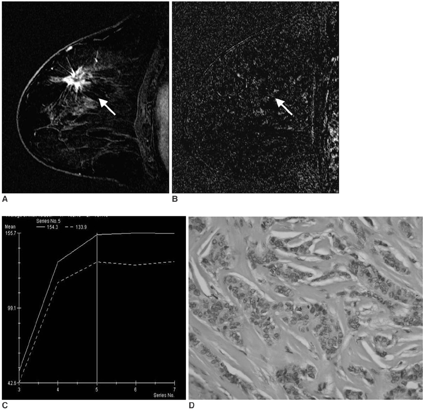
Fig. 1 A 56-year-old woman with invasive ductal carcinoma not otherwise specified of histological grade 1, ER (+), PR (+), Ki-67 (-). A. Sagittal standard subtracted fast lowangle shot MR image shows an irregular mass with a spiculated margin (arrow). B. Sagittal reverse subtracted fast lowangle shot MR image shows nonwashout kinetics (arrow). C. Time-signal intensity curve shows plateau late enhancement. D. Photomicrograph shows high proportional tubule formation, low nuclear pleomorphism, and a low mitotic count (Hematoxylin & Eosin staining, ×400).
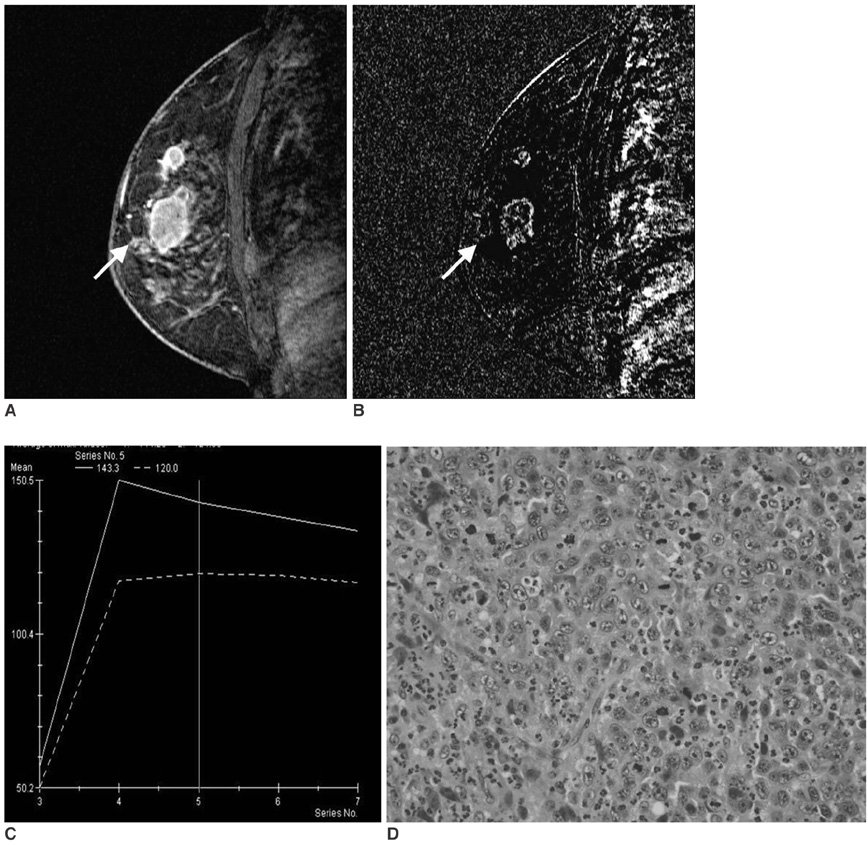
Fig. 2 A 48-year-old woman with invasive ductal carcinoma not otherwise specified of histological grade 3, ER (-), PR (-), Ki-67 (+). A. Sagittal standard subtracted fast lowangle shot MR image shows a lobulated mass with rim enhancement (arrow). B. Sagittal reverse subtracted fast lowangle shot MR image shows washout kinetics (arrow). C. Time-signal intensity curve shows washout late enhancement. D. Photomicrograph shows minimal tubule formation, high nuclear pleomorphism, and a high mitotic count (Hematoxylin & Eosin staining, ×400).
Cited by 2 articles
-
Association between BRCA Mutation Status, Pathological Findings, and Magnetic Resonance Imaging Features in Patients with Breast Cancer at Risk for the Mutation
Jae Myoung Noh, Boo-Kyung Han, Doo Ho Choi, Sun Jung Rhee, Eun Yoon Cho, Seung Jae Huh, Won Park, Hyojung Park, Seok Jin Nam, Jeong Eon Lee, Won-Ho Kil
J Breast Cancer. 2013;16(3):308-314. doi: 10.4048/jbc.2013.16.3.308.Kinetic Features of Invasive Breast Cancers on Computer-Aided Diagnosis Using 3T MRI Data: Correlation with Clinical and Pathologic Prognostic Factors
Sung Eun Song, Kyu Ran Cho, Bo Kyoung Seo, Ok Hee Woo, Seung Pil Jung, Deuk Jae Sung
Korean J Radiol. 2019;20(3):411-421. doi: 10.3348/kjr.2018.0587.
Reference
-
1. Kuhl CK, Mielcareck P, Klaschik S, Leutner C, Wardelmann E, Gieseke J, et al. Dynamic breast MR imaging: are signal intensity time course data useful for differential diagnosis of enhancing lesions? Radiology. 1999. 211:101–110.2. Orel SG. Differentiating benign from malignant enhancing lesions identified at MR imaging of the breast: are time-signal intensity curves an accurate predictor? Radiology. 1999. 211:5–7.3. Mussurakis S, Gibbs P, Horsman A. Peripheral enhancement and spatial contrast uptake heterogeneity of primary breast tumors: quantitative assessment with dynamic MRI. J Comput Assist Tomogr. 1998. 22:35–46.4. Szabó BK, Aspelin P, Kristoffersen Wiberg M, Tot T, Bone B. Invasive breast cancer: correlation of dynamic MR features with prognostic factors. Eur Radiol. 2003. 13:2425–2435.5. Jinguji J, Kajiya Y, Kamimura K, Nakajo M, Sagara Y, Takahama T, et al. Rim enhancement of breast cancers on contrast-enhanced MR imaging: relationship with prognostic factors. Breast Cancer. 2006. 13:64–73.6. Teifke A, Behr O, Schmidt M, Victor A, Vomweg TW, Thelen M, et al. Dynamic MR imaging of breast lesions: correlation with microvessel distribution pattern and histologic characteristics of prognosis. Radiology. 2006. 239:351–360.7. Mussurakis S, Buckley DL, Horsman A. Dynamic MR imaging of invasive breast cancer: correlation with tumor grade and other histologic factors. Br J Radiol. 1997. 70:446–451.8. Bone B, Aspelin P, Bronge L, Veress B. Contrast-enhanced MR imaging as a prognostic indicator of breast cancer. Acta Radiol. 1998. 39:279–284.9. Stomper PC, Herman S, Klippenstein DL, Winston JS, Edge SB, Arredondo MA, et al. Suspect breast lesions: findings at dynamic gadolinium-enhanced MR imaging correlated with mammographic and pathologic features. Radiology. 1995. 197:387–395.10. Fischer U, Kopka L, Brinck U, Korabiowska M, Schauer A, Grabbe E. Prognostic value of contrast-enhanced MR mammography in patients with breast cancer. Eur Radiol. 1997. 7:1002–1005.11. Tuncbilek N, Karakas HM, Okten OO. Dynamic magnetic resonance imaging in determining histopathological prognostic factors of invasive breast cancers. Eur J Radiol. 2005. 53:199–205.12. American College of Radiology. Breast Imaging Reporting and Data System- Magnetic Resonance Imaging. 2003. 1st ed. Reston, VA: American College of Radiology.13. Lee SH, Cho N, Chung HK, Kim SJ, Cha JH, Cho KS, et al. Breast MR imaging: correlation of high resolution dynamic MR findings with prognostic factors. J Korean Radiol Soc. 2005. 52:355–361. (Korean).14. Elston CW, Ellis IO, Pinder SE. Pathologic prognostic factors in breast cancer. Crit Rev Oncol Hematol. 1999. 31:209–223.15. Elston CW, Ellis IO. Pathological prognostic factors in breast cancer. I. The value of histological grade in breast cancer: experience from a large study with long-term follow-up. Histopathology. 1991. 19:403–410.16. Stavros AT, Thickman D, Rapp CL, Dennis MA, Parker SH, Sisney GA. Solid breast nodule: use of sonography to distinguish between benign and malignant lesions. Radiology. 1995. 196:123–134.17. Stavros AT. Stavros AT, editor. Malignant solid breast nodules: specific type. Breast ultrasound. 2004. 1st ed. Philadelphia: Lippincott Williams & Wilkins;597–688.18. Buadu LD, Murakami J, Murayama S, Hashiguchi N, Sakai S, Masuda K, et al. Breast lesions: correlation of contrast medium enhancement patterns on MR images with histopathologic findings and tumor angiogenesis. Radiology. 1996. 200:639–649.19. Buckley DL, Drew PJ, Mussurakis S, Monson JR, Horsman A. Microvessel density of invasive breast cancer assessed by dynamic Gd-DTPA enhanced MRI. J Magn Reson Imaging. 1997. 7:461–464.20. Matsubayashi R, Matsuo Y, Edakuni G, Satoh T, Tokunaga O, Kudo S. Breast masses with peripheral rim enhancement on dynamic contrast-enhanced MR images: correlation of MR findings with histologic features and expression of growth factors. Radiology. 2000. 217:841–848.21. Stomper PC, Herman S, Klippenstein DL, Winston JS, Budnick RM, Stewart CC. Invasive breast carcinoma: analysis of dynamic magnetic resonance imaging enhancement features and cell proliferative activity determined by DNA S-phase percentage. Cancer. 1996. 77:1844–1849.22. Heywang-Kobrunner SH. Contrast enhanced magnetic resonance imaging of the breast. Invest Radiol. 1994. 29:94–104.
- Full Text Links
-
- Actions
-
Cited
- CITED
-
- Close
- Share
-
- Similar articles
-
- Breast MR Imaging: Correlation of High Resolution Dynamic MR Findings with Prognostic Factors
- Dynamic MRI for Breast Cancer: Correlation with the Prognostic factors and the Time-Signal Intensity Curve
- Dynamic MRI of Breast Fibroadenoma: Pathologic Correlation
- Prognostic Implications of MicroRNA-21 Overexpression in Invasive Ductal Carcinomas of the Breast
- Comments on the "Prognostic Impact and Clinicopathological Correlation of CD133 and ALDH1 Expression in Invasive Breast Cancer"
