Korean J Radiol.
2010 Oct;11(5):571-573. 10.3348/kjr.2010.11.5.571.
Linear Fat Deposition in the Middle Layer of the Left Ventricular Myocardium: Computed Tomographic Findings
- Affiliations
-
- 1Department of Radiology, Chungnam National University Hospital, Chungnam National University School of Medicine, Daejeon 301-721, Korea.
- 2Department of Radiology, Konkuk University Hospital, Konkuk University School of Medicine, Seoul 143-729, Korea. ksm9723@yahoo.co.kr
- 3Department of Thoracic Surgery, Konkuk University Hospital, Konkuk University School of Medicine, Seoul 143-729, Korea.
- KMID: 1102584
- DOI: http://doi.org/10.3348/kjr.2010.11.5.571
Abstract
- We report here a case of streaky fat deposition in the middle layer of the left ventricular myocardium, without any underlying etiology, and this was seen on computed tomography coronary angiography. This report suggests that left ventricular middle layer fat deposition should be investigated in order to determine its etiology, the pathogenesis and the prognosis.
Keyword
MeSH Terms
Figure
-
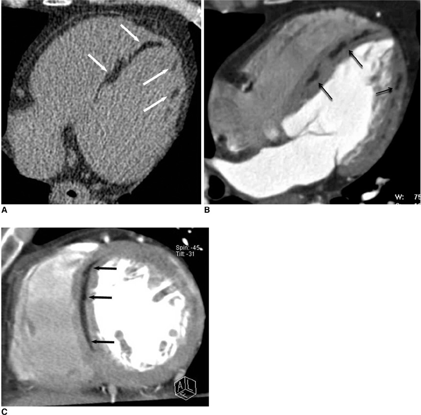
Fig. 1 Axial unenhanced and contrast-enhanced CT scans (A, B) show curvilinear and patchy foci of fat involving septum and lateral wall of left ventricle. Short-axis contrast-enhanced CT scan (C) demonstrates fat streak (arrows) within hypertrophied mid septum of left ventricular myocardium. There was no stenosis in right or left coronary arteries (not shown).
Cited by 1 articles
-
Assessment of Left Ventricular Myocardial Diseases with Cardiac Computed Tomography
Sung Min Ko, Tae Hoon Kim, Eun Ju Chun, Jin Young Kim, Sung Ho Hwang
Korean J Radiol. 2019;20(3):333-351. doi: 10.3348/kjr.2018.0280.
Reference
-
1. Fontaine G, Fontaliran F, Zenati O, Guzman CE, Rigoulet J, Berthier JL, et al. Fat in the heart. A feature unique to the human species? Observational reflections on an unsolved problem. Acta Cardiol. 1999. 54:189–194.2. Tandri H, Bomma C, Calkins H, Bluemke DA. Magnetic resonance and computed tomography imaging of arrhythmogenic right ventricular dysplasia. J Magn Reson Imaging. 2004. 19:848–858.3. Gaerte SC, Meyer CA, Winer-Muram HT, Tarver RD, Conces DJ Jr. Fat-containing lesions of the chest. Radiographics. 2002. 22:S61–S78.4. Heyer CM, Kagel T, Lemburg SP, Bauer TT, Nicolas V. Lipomatous hypertrophy of the interatrial septum: a prospective study of incidence, imaging findings, and clinical symptoms. Chest. 2003. 124:2068–2073.5. Kaminaga T, Naito H, Takamiya M, Hamada S, Nishimura T. Myocardial damage in patients with dilated cardiomyopathy: CT evaluation. J Comput Assist Tomogr. 1994. 18:393–397.6. Ahn SS, Kim YJ, Hur J, Lee HJ, Kim TH, Choe KO, et al. CT detection of subendocardial fat in myocardial infarction. AJR Am J Roentgenol. 2009. 192:532–537.7. Zafar HM, Litt HI, Torigian DA. CT imaging features and frequency of left ventricular myocardial fat in patients with CT findings of chronic left ventricular myocardial infarction. Clin Radiol. 2008. 63:256–262.8. Kimura F, Sakai F, Sakomura Y, Fujimura M, Ueno E, Matsuda N, et al. Helical CT features of arrhythmogenic right ventricular cardiomyopathy. Radiographics. 2002. 22:1111–1124.9. Kaminaga T, Naitou H, Hamada S, Takamiya M. Detection of myocardial fatty components with ultrafast CT. Nippon Igaku Hoshasen Gakkai Zasshi. 1993. 53:28–34. [Japanese].10. Carpenter HM. Myocardial fat infiltration. Am Heart J. 1962. 63:491–496.
- Full Text Links
-
- Actions
-
Cited
- CITED
-
- Close
- Share
-
- Similar articles
-
- Fat Deposition in the Urinary Bladder Wall: Incidental Finding on Abdominal Computed Tomography: A Case Report
- Noncompaction of Ventricular Myocardium Involving the Right Ventricle
- A Case of Noncompaction of the Ventricular Myocardium Combined with Situs Ambiguous with Polysplenia
- Stroke in a Young Individual with Left Ventricular Noncompaction and Left Atrium Standstill
- Three-Dimensional Endo-Cardiovascular Volume-Rendered Cine Computed Tomography of Isolated Left Ventricular Apical Hypoplasia: A Case Report and Literature Review
