Three-Dimensional Endo-Cardiovascular Volume-Rendered Cine Computed Tomography of Isolated Left Ventricular Apical Hypoplasia: A Case Report and Literature Review
- Affiliations
-
- 1Department of Radiology, Sejong General Hospital, Bucheon 14754, Korea. ymkimna@naver.com
- 2Department of Internal Medicine, Division of Cardiology, Sejong General Hospital, Bucheon 14754, Korea.
- KMID: 2351166
- DOI: http://doi.org/10.3348/kjr.2016.17.1.79
Abstract
- We report multidetector computed tomography (MDCT) and cardiac magnetic resonance (CMR) findings of a 34-year-old female with isolated left ventricular apical hypoplasia. The MDCT and CMR scans displayed a spherical left ventricle (LV) with extensive fatty infiltration within the myocardium at the apex, interventricular septum and inferior wall, anteroapical origin of the papillary muscle, right ventricle wrapping around the deficient LV apex, and impaired systolic function. MDCT visualized morphologic and also functional findings of this unique cardiomyopathy.
Keyword
MeSH Terms
Figure
-
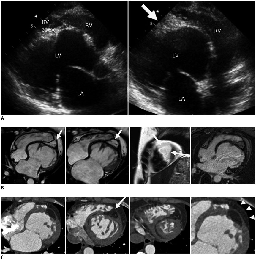
Fig. 1 Isolated left ventricular apical hypoplasia in 34-year-old woman. A. Echocardiogram demonstrates spherical and truncated LV with echo-bright apex (arrow), bulging of interventricular septum towards right ventricle, and elongated RV wrapping around deficient LV apex in parasternal long-axis view. B. Four-chamber view from CMR during diastole and systole shows spherical, truncated LV at apical level with prominent fat (arrows). Papillary muscles originate from flattened anterior apex. RV is elongated and wraps around deficient LV apex. Late gadolinium enhancement revealed no enhancement in apex and ventricular septum of LV. C. Four-chamber view from cardiac MDCT more clearly visualizes extent of fatty material within apical myocardium, interventricular septum and inferior LV wall. Almost all fatty material was confined within myocardium, although small portion of apical fat was contiguous with epicardial fat (arrow). MDCT clearly shows cardiac layers and fat distribution. Fatty tissue is confined within thin myocardial layers of apex and interventricular septum. Note pericardium (arrowheads) and epicardial fat. CMR = cardiac magnetic resonance, LA = left atrium, LV = left ventricle, MDCT = multidetector computed tomography, RV = right ventricle, VR = volume-rendering
Cited by 1 articles
-
User-Friendly Vendor-Specific Guideline for Pediatric Cardiothoracic Computed Tomography Provided by the Asian Society of Cardiovascular Imaging Congenital Heart Disease Study Group: Part 1. Imaging Techniques
Sun Hwa Hong, Hyun Woo Goo, Eriko Maeda, Ki Seok Choo, I-Chen Tsai,
Korean J Radiol. 2019;20(2):190-204. doi: 10.3348/kjr.2018.0571.
Reference
-
1. Fernandez-Valls M, Srichai MB, Stillman AE, White RD. Isolated left ventricular apical hypoplasia: a new congenital anomaly described with cardiac tomography. Heart. 2004; 90:552–555.2. Marin C, Sanchez ML, Maroto E, Ossaba S, Ruiz Y, Zabala JI. MR imaging of isolated left ventricular apical hypoplasia. Pediatr Radiol. 2007; 37:703–705.3. Flett AS, Elliott PM, Moon JC. Images in cardiovascular medicine. Cardiovascular magnetic resonance of isolated left ventricular apical hypoplasia. Circulation. 2008; 117:e504–e505.4. Irving CA, Chaudhari MP. Fatal presentation of congenital isolated left ventricular apical hypoplasia. Eur J Cardiothorac Surg. 2009; 35:368–369.5. Patrianakos AP, Protonotarios N, Zacharaki A, Tsatsopoulou A, Parthenakis FI, Vardas PE. Isolated left ventricular apical hypoplasia: a newly recognized unclassified cardiomyopathy. J Am Soc Echocardiogr. 2010; 23:1336.e1–1336.e4.6. Haffajee JA, Finley JJ, Brooks EL, Kuvin JT, Patel AR. Echocardiographic characterization of left ventricular apical hypoplasia accompanied by a patent ductus arteriosus. Eur J Echocardiogr. 2011; 12:E17.7. Moon JI, Jeong YJ, Lee G, Choi JH, Lee JW. Isolated left ventricular apical hypoplasia with infundibular pulmonary and aortic stenosis: a rare combination. Korean J Radiol. 2013; 14:874–877.8. Chaowu Y, Xin S, Shihua Z, Jianrong L, Hao W. Complete transposition of the atrioventricular valves associated with left ventricular apical hypoplasia. Circulation. 2011; 124:e538–e539.9. Motwani M, Witte KK, Plein S, Greenwood JP. Isolated left ventricular apical hypoplasia evaluated by cardiovascular magnetic resonance and gadolinium enhancement techniques. J Am Coll Cardiol. 2011; 58:2355.10. Francone M. Role of cardiac magnetic resonance in the evaluation of dilated cardiomyopathy: diagnostic contribution and prognostic significance. ISRN Radiol. 2014; 2014:365404.11. Coats CJ, Quarta G, Flett AS, Pantazis AA, McKenna WJ, Moon JC. Arrhythmogenic left ventricular cardiomyopathy. Circulation. 2009; 120:2613–2614.12. Paetsch I, Reith S, Gassler N, Jahnke C. Isolated arrhythmogenic left ventricular cardiomyopathy identified by cardiac magnetic resonance imaging. Eur Heart J. 2011; 32:2840.
- Full Text Links
-
- Actions
-
Cited
- CITED
-
- Close
- Share
-
- Similar articles
-
- Isolated Left Ventricular Apical Hypoplasia with Infundibular Pulmonary and Aortic Stenosis: a Rare Combination
- Comparison of Left and Right Ventricular Volume and Cardiac Output by MRI and Echocardiography
- Comparison between Echocardiography and Cardiac Cine-MRI : Left Ventricular Volume and Cardiac Output
- Isolated Right Ventricular Apical Hypoplasia: A Case Report with 18 Years of Follow Up
- Isolated Right Ventricular Hypoplasia: A case report
