Video-Assisted Thoracoscopic Surgery for Correction of Adolescent Idiopatic Scoliosis: Comparison of 4.5 mm versus 5.5 mm Rod Constructs
- Affiliations
-
- 1Department of Orthopaedic Surgery, Yonsei University College of Medicine, Seoul, Korea. mes1007@yuhs.ac
- 2Department of Orthopaedic Surgery, National Health Insurance Corporation Ilsan Hospital, Goyang, Korea.
- 3Department of Anesthesiology and Pain Medicine, National Health Insurance Corporation Ilsan Hospital, Goyang, Korea.
- 4Department of Orthopaedic Surgery, CHA University, Pocheon, Korea.
- 5Department of Radiology, Yonsei University College of Medicine, Seoul, Korea.
- KMID: 1071429
- DOI: http://doi.org/10.3349/ymj.2010.51.5.753
Abstract
- PURPOSE
The purpose of this study is to report the comparative results of thoracoscopic correction achieved via cantilever technique using a 4.5 mm thin rod and the poly-axial reduction screw technique using a 5.5 mm thick rod in Lenke type 1 adolescent idiopathic scoliosis (AIS).
MATERIALS AND METHODS
Radiographic data, Scoliosis Research Society (SRS) patient-based outcome questionnaires, and operative records were reviewed for forty-nine patients undergoing surgical treatment of scoliosis. The study group was divided into a 4.5 mm thin rod group (n = 24) and a 5.5 mm thick rod group (n = 25). The radiographic parameters that were analyzed included coronal curve correction, the most caudal instrumented vertebra tilt angle correction, coronal balance, and thoracic kyphosis.
RESULTS
The major curve was corrected from 49.8degrees and 47.2degrees pre-operatively to 24.5degrees and 18.8degrees at the final follow-up for the thin and thick rod groups, respectively (50.8% vs. 60.2% correction). There were no significant differences between the two groups in terms of kyphosis, coronal balance, or tilt angle at the time of the final follow-up. The mean number of levels fused was 6.2 in the thin rod group, compared with 5.9 levels in the thick rod group. There were no major intraoperative complications in either group.
CONCLUSION
Significant correction loss was observed in the thin rod system at the final follow-up though both groups had comparable correction immediately post-operative. Therefore, the thick rod with poly axial screw system helps to maintain post-operative correction.
Keyword
MeSH Terms
Figure
-
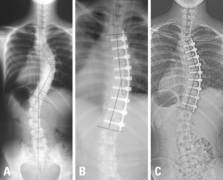
Fig. 1 Radiographs of the spine in a patient instrumented with thin 4.5 mm diameter rod. (A) Pre-operative anteroposterior radiograph. (B) Immediate post-operative anteroposterior supine radiograph. (C) The anteroposterior standing radiograph made thirty months after surgery, showing the large degree correction loss compared to immediate post-operative radiograph.
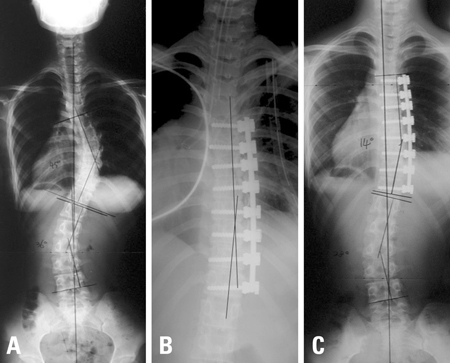
Fig. 2 Radiographs of the spine in a patient instrumented with thick 5.5 mm diameter rod. (A) Pre-operative anteroposterior radiograph. (B) Immediate post-operative anteroposterior supine radiograph. (C) The anteroposterior standing radiograph made thirty-eight months after surgery, showing no loss of correction compared to immediate post-operative radiograph.
Cited by 1 articles
-
Video-Assisted Thoracoscopic Surgery Plus Lumbar Mini-Open Surgery for Adolescent Idiopathic Scoliosis
Hyon Su Chong, Hak Sun Kim, Nanda Ankur, Phillip Anthony Kho, Sung Jun Kim, Do Yeon Kim, Jin Oh Park, Seong Hwan Moon, Hwan Mo Lee, Eun Su Moon
Yonsei Med J. 2011;52(1):130-136. doi: 10.3349/ymj.2011.52.1.130.
Reference
-
1. Karami M, Ilharreborde B, Morel E, Fitoussi F, Penneçot GF, Mazda K. Video-assisted thoracoscopic surgery (VATS) for the treatment of scolioticrib hump deformity. Eur Spine J. 2007. 16:1373–1377.
Article2. Kim HS, Lee CS, Jeon BH, Park JO. Sagittal plane analysis of adolescent idiopathic scoliosis after VATS (video-assisted thoracoscopic surgery) anterior instrumentations. Yonsei Med J. 2007. 48:90–96.
Article3. Lenke LG, Newton PO, Marks MC, Blanke KM, Sides B, Kim YJ, et al. Prospective pulmonary function comparison of open versus endoscopic anterior fusion combined with posterior fusion in adolescent idiopathic scoliosis. Spine (Phila Pa 1976). 2004. 29:2055–2060.
Article4. Lonner BS, Kondrachov D, Siddiqi F, Hayes V, Scharf C. Thoracoscopic spinal fusion compared with posterior spinal fusion for the treatment of thoracic adolescent idiopathic scoliosis. Surgical technique. J Bone Joint Surg Am. 2007. 89(Pt. 1):Suppl 2. 142–156.
Article5. Newton PO, Marks M, Faro F, Betz R, Clements D, Haher T, et al. Use of video-assisted thoracoscopic surgery to reduce perioperative morbidity in scoliosis surgery. Spine (Phila Pa 1976). 2003. 28:S249–S254.
Article6. Wong HK, Hee HT, Yu Z, Wong D. Results of thoracoscopic instrumented fusion versus conventional posterior instrumented fusion in adolescent idiopathic scoliosis undergoing selective thoracic fusion. Spine (Phila Pa 1976). 2004. 29:2031–2038.
Article7. Crawford AH, Wall EJ, Wolf R. Video-assisted thoracoscopy. Orthop Clin North Am. 1999. 30:367–385.
Article8. Ohtsuka T, Ohnishi I, Nakamura K, Takamoto S. New instrumentation for video-assisted anterior spine release. Surg Endosc. 2000. 14:682–684.9. Betz RR, Harms J, Clements DH 3rd, Lenke LG, Lowe TG, Shufflebarger HL, et al. Comparison of anterior and posterior instrumentation for correction of adolescent thoracic idiopathic scoliosis. Spine (Phila Pa 1976). 1999. 24:225–239.
Article10. Sweet FA, Lenke LG, Bridwell KH, Blanke KM, Whorton J. Prospective radiographic and clinical outcomes and complications of single solid rod instrumented anterior spinal fusion in adolescent idiopathic scoliosis. Spine (Phila Pa 1976). 2001. 26:1956–1965.
Article11. Newton PO, Parent S, Marks M, Pawelek J. Prospective evaluation of 50 consecutive scoliosis patients surgically treated with thoracoscopic anterior instrumentation. Spine (Phila Pa 1976). 2005. 30:S100–S109.
Article12. Lenke LG, Betz RR, Harms J, Bridwell KH, Clements DH, Lowe TG, et al. Adolescent idiopathic scoliosis: a new classification to determine extent of spinal arthrodesis. J Bone Joint Surg Am. 2001. 83-A:1169–1181.13. Lonner BS, Kondrachov D, Siddiqi F, Hayes V, Scharf C. Thoracoscopic spinal fusion compared with posterior spinal fusion for the treatment of thoracic adolescent idiopathic scoliosis. J Bone Joint Surg Am. 2006. 88:1022–1034.
Article14. Picetti GD 3rd, Ertl JP, Bueff HU. Endoscopic instrumentation, correction, and fusion of idiopathic scoliosis. Spine J. 2001. 1:190–197.
Article15. Son-Hing JP, Blakemore LC, Poe-Kochert C, Thompson GH. Video-assisted thoracoscopic surgery in idiopathic scoliosis: evaluation of the learning curve. Spine (Phila Pa 1976). 2007. 32:703–707.16. Norton RP, Patel D, Kurd MF, Picetti GD, Vaccaro AR. The use of thoracoscopy in the management of adolescent idiopathic scoliosis. Spine (Phila Pa 1976). 2007. 32:2777–2785.
Article17. Harms J, Jeszenszky D, Beele B. Bridwell K, DeWald R, editors. Ventral correction of thoracic scoliosis. The textbook of spinal surgery. 1997. Vol 40. Philedelphia: Lipincott-Raven;611–626.18. Lenke LG, Betz RR, Bridwell KH, Harms J, Clements DH, Lowe TG. Spontaneous lumbar curve coronal correction after selective anterior or posterior thoracic fusion in adolescent idiopathic scoliosis. Spine (Phila Pa 1976). 1999. 24:1663–1671.
Article19. Polly DW Jr, Cunningham BW, Kuklo TR, Lenke LG, Oda I, Schroeder TM, et al. Anterior thoracic scoliosis constructs: effect of rod diameter and intervertebral cages on multi-segmental construct stability. Spine J. 2003. 3:213–219.20. Wedemeyer M, Parent S, Mahar A, Odell T, Swimmer T, Newton P. Titanium versus stainless steel for anterior spinal fusions: an analysis of rod stress as a predictor of rod breakage during physiologic loading in a bovine model. Spine (Phila Pa 1976). 2007. 32:42–48.21. Yoon SH, Ugrinow VL, Upasani VV, Pawelek JB, Newton PO. Comparison between 4.0-mm stainless steel and 4.75-mm titanium alloy single-rod spinal instrumentation for anterior thoracoscopic scoliosis surgery. Spine (Phila Pa 1976). 2008. 33:2173–2178.
Article22. Asher MA, Min Lai S, Burton DC. Further development and validation of the Scoliosis Research Society (SRS) outcomes instrument. Spine (Phila Pa 1976). 2000. 25:2381–2386.
Article23. Liljenqvist UR, Bullmann V, Schulte TL, Hackenberg L, Halm HF. Anterior dual rod instrumentation in idiopathic thoracic scoliosis. Eur Spine J. 2006. 15:1118–1127.
Article24. Climent JM, Reig A, Sáhcnez J, Roda C. Construction and validation of a specific quality of life instrument for adolescents with spine deformities. Spine (Phila Pa 1976). 1995. 20:2006–2011.
Article
- Full Text Links
-
- Actions
-
Cited
- CITED
-
- Close
- Share
-
- Similar articles
-
- Polyaxial Screws with Thick Rod versus Monoaxial Screws with Thin Rod in Video-Assisted Thoracoscopic Scoliosis Surgery (VATS)
- Choice of Rods in Surgical Treatment of Adolescent Idiopathic Scoliosis: What Are the Clinical Implications of Biomechanical Properties? – A Review of the Literature
- Use of Harrington Compression Rod
- A Case of Iatrogenic Horner's Syndrome after Video-Thoracoscopic Surgery for Primary Pneumothorax
- Video-Assisted Thoracoscopic Surgery Plus Lumbar Mini-Open Surgery for Adolescent Idiopathic Scoliosis
