J Korean Orthop Assoc.
1974 Dec;9(4):456-460. 10.4055/jkoa.1974.9.4.456.
Use of Harrington Compression Rod
- Affiliations
-
- 1Department of Orthopedic Surgery, Seoul National University Hospital, Korea.
- KMID: 2471470
- DOI: http://doi.org/10.4055/jkoa.1974.9.4.456
Abstract
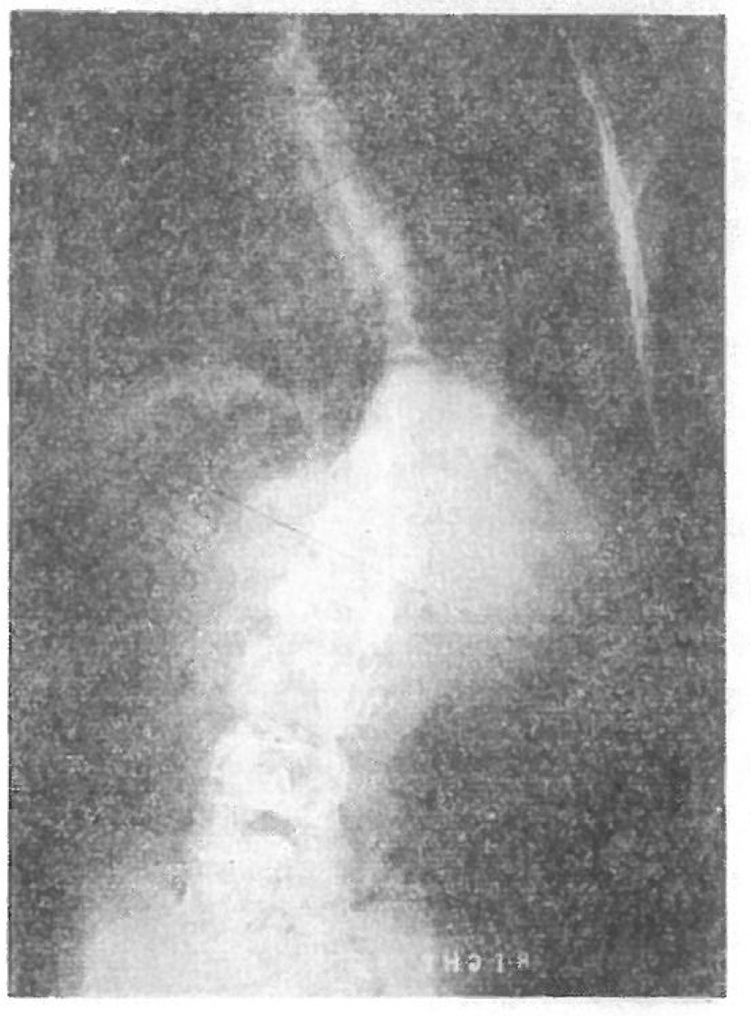
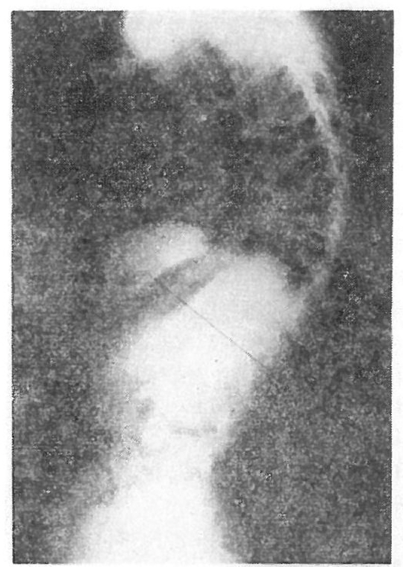
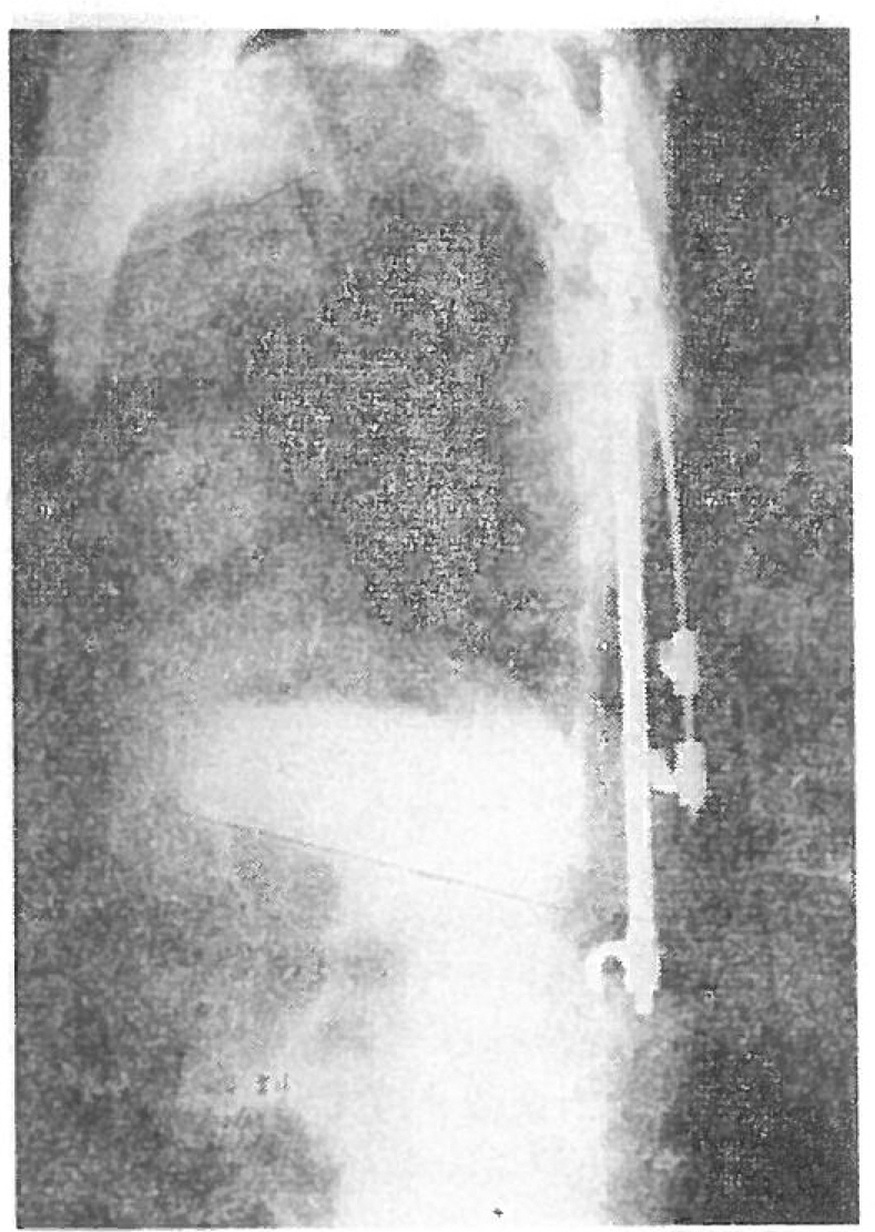
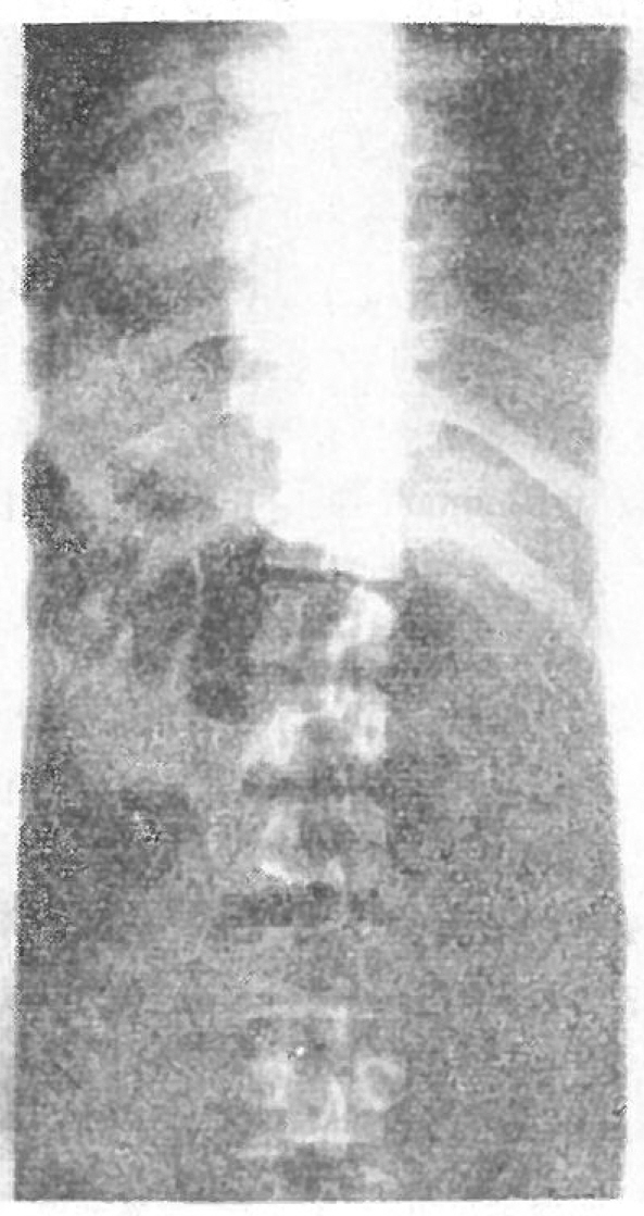
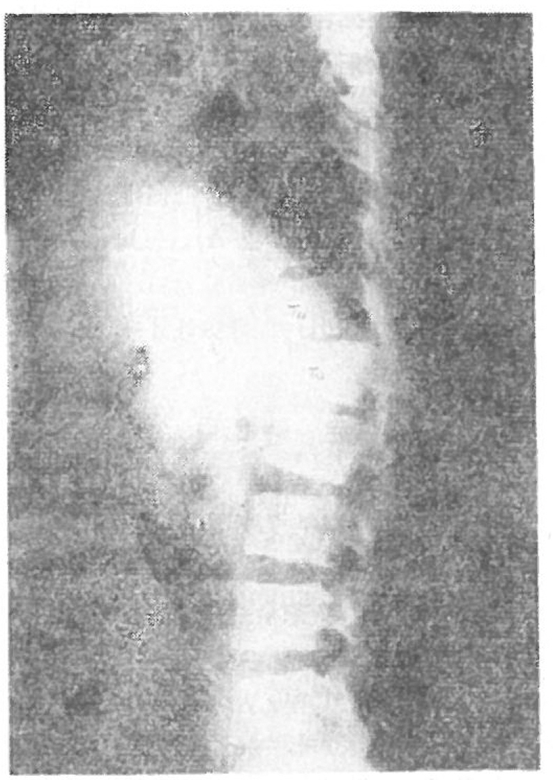
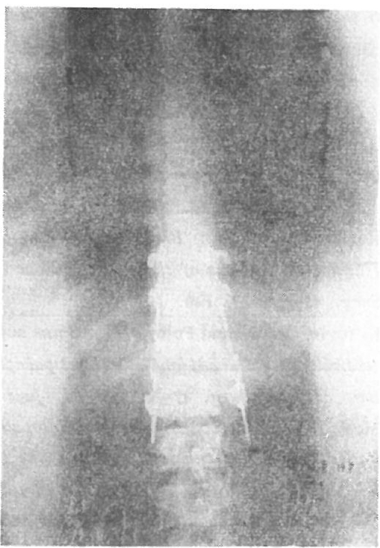
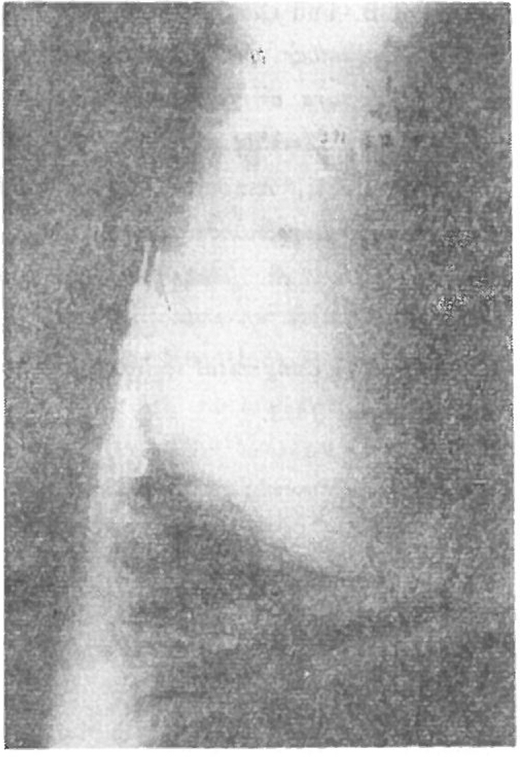
- It depends on orthopedist's opinion whether to use Harrington compression rod for correction of scoliosis with Harrington distraction rod, for example, Dr. Harrington and Hall rocommend it, but Dr. Moe and Goldstein do not use it routinely for simple scoliosis curve. The disadvantage of use of compression rod is first of all prologation of operation time without addition of significant correction of scoliosis. Recently the author used Harrington compression rod for a case of congenital kyphoscoliosis and anterior T11-T12, fracture-dislocation respectively. The result was remarkable correction of kyphotic curve from 73° to 43° and scoliolic curve from 54° to 19°. In the spinal fracture-dislocation case it was felt that the dislocation site was adequately stabilized by bilateral Harrington compression rods which were inserted in the transverse processes of T10 to L2. The purpose of this paper is to alert orthopedist that Harrington compression rod is useful of kyphoscoliosis correction and stabilization of a certain spinal fracture-dislocation.
MeSH Terms
Figure
Reference
-
1. Goldstein L.A.The surgical treatment of idiopathic scoliosis. Clin. Orthop. 93:131–157. June. 1973.
Article2. Hall J.E.Idiopathic scoliosis, indications for treatment and follow-up study of 210 cases treated by Harrington instrumentation. Second Annual Postgraduate Course on the Mangement and Care of the Scoliosis Patient. 23–26. Nov. 1970.3. Harrington P.R.Treatment of scoliosis. Correction and internal fixation by spine instrumentation. J Bone & Joint Surg. 44-A:591–610. June. 1973.4. Harrington P.R., Dickson J.H.An eleven-year clinical investigation of Harrington instrumentation. Clin. Orthop. 93:113–30. June. 1973.5. Holdsworth F.W.Fractures, dislocations and fracture-dislocations of the spine. J Bone & Joint Surg. 45-A:1–20. Feb. 1963.
Article6. Kilfoyle R.M., Foley J.J.Spine and pelvic deformity in childhood and adolescent paraplegia. J Bone & Joinn Surg. 47-A:659–683. June. 1965.
Article7. Moe J.H., Kettleson D.N.Idiopathic scoliosis, Analysis of curve pattern and the preliminary results of Milwaukee Brace treatment in one hundred sixty-nine patients. J Bone and Joint Surg. 52-A:1509–1633. Dec. 1973.8. Moe J.H.Methods of correction and surgical techniques in scoliosis. The Orthop. Clin. of North Am. 3(1):17–48. Mar. 1971.
Article9. Roberts J.B., Curtiss P.H.Stability of the thoracic and lumbar spine in traumatic paraplegia following fracture or fracturedislocation. J Bone & Joint Surg. 52-A:1115–1130. Sept. 1970.
Article10. Tamborino J.M., Amburst E.N., Moe J.H.Harrington instrumentation in correction of scoliosis. J Bone & Joint Surg. 46-A:313–323. Mar. 1964.11. Winter R.B.Congenital scoliosis. Clin. Orthop. 93:75–94. June. 1973.
Article
- Full Text Links
- Actions
-
Cited
- CITED
-
- Close
- Share
- Similar articles
-
- Segmental Spinal Instrumentation in the Management of Fracture and Fracture-Dislocation of the Thoraco-Lumbar Spine
- Clinical Results of Segmental Spinal Instrumentation in Unstable Fracture and Fracture-Dislocation of the Thoracolumbar Spine
- Clinical Analysis of Unstable Thoracolumbar Fracture and Fracture-dislocation Using Transpedicular Screws and Harrington distration rod
- Clinical Results of Segmental spinal instrumentation in Unstable Fracture and Fracture - Dislocation of the Thoracolumbar Spine
- A Clinical Study of the Surgical Treatment of the Thoraco-Lumbar Spinal Injuries

