Yonsei Med J.
2011 Jan;52(1):130-136. 10.3349/ymj.2011.52.1.130.
Video-Assisted Thoracoscopic Surgery Plus Lumbar Mini-Open Surgery for Adolescent Idiopathic Scoliosis
- Affiliations
-
- 1Department of Orthopaedic Surgery, Yonsei University College of Medicine, Seoul, Korea. mes1007@yuhs.ac
- 2Indian Spinal injuries Centre, New Delhi, India.
- 3Department of Radiology, Yonsei University College of Medicine, Seoul, Korea.
- KMID: 1106447
- DOI: http://doi.org/10.3349/ymj.2011.52.1.130
Abstract
- PURPOSE
The objectives of this study are to describe the outcome of adolescent idiopathic scoliosis (AIS) patients treated with Video Assisted Thoracoscopic Surgery (VATS) plus supplementary minimal incision in the lumbar region for thoracic and lumbar deformity correction and fusion.
MATERIALS AND METHODS
This is a case series of 13 patients treated with VATS plus lumbar mini-open surgery for AIS. A total of 13 patients requiring fusions of both the thoracic and lumbar regions were included in this study: 5 of these patients were classified as Lenke type 1A and 8 as Lenke type 5C. Fusion was performed using VATS up to T12 or L1 vertebral level. Lower levels were accessed via a small mini-incision in the lumbar area to gain access to the lumbar spine via the retroperitoneal space. All patients had a minimum follow-up of 1 year.
RESULTS
The average number of fused vertebrae was 7.1 levels. A significant correction in the Cobb angle was obtained at the final follow-up (p = 0.001). The instrumented segmental angle in the sagittal plane was relatively well-maintained following surgery, albeit with a slight increase. Scoliosis Research Society-22 (SRS-22) scores were noted have significantly improved at the final follow-up (p < 0.05).
CONCLUSION
Indications for the use of VATS may be extended from patients with localized thoracic scoliosis to those with thoracolumbar scoliosis. By utilizing a supplementary minimal incision in the lumbar region, a satisfactory deformity correction may be accomplished with minimal post-operative scarring.
Keyword
MeSH Terms
Figure
-
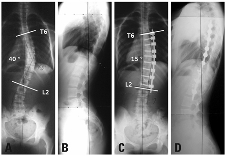
Fig. 1 Preoperative posteroanterior (A) and lateral (B) radiograph of 12 year-old girl with 40O T6-L2 Lenke type 1A curve. The post-operative 2 year follow-up posteroanterior (C) and lateral (D) radiograph of same patient after VATS and lumbar mini-open surgery and fusion. VATS, Video Assisted Thoracoscopic Surgery.

Fig. 2 Preoperative posteroanterior (A) and lateral (B) radiograph of 14 year-old girl with 44O T10-L3 Lenke type 5C curve. The post-operative 3 year follow-up posteroanterior (C) and lateral (D) radiograph of same patient after VATS and lumbar mini-open surgery and fusion. VATS, Video Assisted Thoracoscopic Surgery.
Reference
-
1. Weinstein SL. Adolescent idiopathic scoliosis: prevalence and natural history. Instr Course Lect. 1989. 38:115–128.2. Noonan KJ, Dolan LA, Jacobson WC, Weinstein SL. Long-term psychosocial characteristics of patients treated for idiopathic scoliosis. J Pediatr Orthop. 1997. 17:712–717.
Article3. Picetti GD 3rd, Ertl JP, Bueff HU. Anterior endoscopic correction of scoliosis. Orthop Clin North Am. 2002. 33:421–429.
Article4. Bernstein RM, Hall JE. Solid rod short segment anterior fusion in thoracolumbar scoliosis. J Pediatr Orthop B. 1998. 7:124–131.
Article5. Landreneau RJ, Hazelrigg SR, Mack MJ, Dowling RD, Burke D, Gavlick J, et al. Postoperative pain-related morbidity: video-assisted thoracic surgery versus thoracotomy. Ann Thorac Surg. 1993. 56:1285–1289.6. Newton PO, Marks M, Faro F, Betz R, Clements D, Haher T, et al. Use of video-assisted thoracoscopic surgery to reduce perioperative morbidity in scoliosis surgery. Spine (Phila Pa 1976). 2003. 28:S249–S254.
Article7. Kim HS, Park JO, Nanda A, Kho PA, Kim JY, Lee HM, et al. Video-assisted thoracoscopic surgery for correction of adolescent idiopatic scoliosis: comparison of 4.5 mm versus 5.5 mm rod constructs. Yonsei Med J. 2010. 51:753–760.
Article8. Brodner W, Mun Yue W, Möller HB, Hendricks KJ, Burd TA, Gaines RW. Short segment bone-on-bone instrumentation for single curve idiopathic scoliosis. Spine (Phila Pa 1976). 2003. 28:S224–S233.
Article9. Betz RR, Shufflebarger H. Anterior versus posterior instrumentation for the correction of thoracic idiopathic scoliosis. Spine (Phila Pa 1976). 2001. 26:1095–1100.
Article10. Burton DC, Asher MA, Lai SM. Patient-based outcomes analysis of patients with single torsion thoracolumbar-lumbar scoliosis treated with anterior or posterior instrumentation: an average 5- to 9-year follow-up study. Spine (Phila Pa 1976). 2002. 27:2363–2367.
Article11. Suk SI, Lee CK, Chung SS. Comparison of Zielke ventral derotation system and Cotrel-Dubousset instrumentation in the treatment of idiopathic lumbar and thoracolumbar scoliosis. Spine (Phila Pa 1976). 1994. 19:419–429.12. Cochran T, Irstam L, Nachemson A. Long-term anatomic and functional changes in patients with adolescent idiopathic scoliosis treated by Harrington rod fusion. Spine (Phila Pa 1976). 1983. 8:576–584.
Article13. Hee HT, Yu ZR, Wong HK. Comparison of segmental pedicle screw instrumentation versus anterior instrumentation in adolescent idiopathic thoracolumbar and lumbar scoliosis. Spine (Phila Pa 1976). 2007. 32:1533–1542.
Article14. Luk KD, Leong JC, Reyes L, Hsu LC. The comparative results of treatment in idiopathic thoracolumbar and lumbar scoliosis using the Harrington, Dwyer, and Zielke instrumentation. Spine (Phila Pa 1976). 1989. 14:275–280.
Article15. Lowe TG, Peters JD. Anterior spinal fusion with Zielke instrumentation for idiopathic scoliosis. A frontal and sagittal curve analysis in 36 patients. Spine (Phila Pa 1976). 1993. 18:423–426.
Article16. Moskowitz A, Trommanhauser S. Surgical and clinical results of scoliosis surgery using Zielke instrumentation. Spine (Phila Pa 1976). 1993. 18:2444–2451.
Article17. Kostuik JP, Carl A, Ferron S. Anterior Zielke instrumentation for spinal deformity in adults. J Bone Joint Surg Am. 1989. 71:898–912.
Article18. Landreneau RJ, Hazelrigg SR, Mack MJ, Dowling RD, Burke D, Gavlick J, et al. Postoperative pain-related morbidity: video-assisted thoracic surgery versus thoracotomy. Ann Thorac Surg. 1993. 56:1285–1289.
Article19. Pollock ME, O'Neal K, Picetti G, Blackman R. Results of video-assisted exposure of the anterior thoracic spine in idiopathic scoliosis. Ann Thorac Surg. 1996. 62:818–823.
Article20. Newton PO, Wenger DR, Mubarak SJ, Meyer RS. Anterior release and fusion in pediatric spinal deformity. A comparison of early outcome and cost of thoracoscopic and open thoracotomy approaches. Spine (Phila Pa 1976). 1997. 22:1398–1406.21. Newton PO, Cardelia JM, Farnsworth CL, Baker KJ, Bronson DG. A biomechanical comparison of open and thoracoscopic anterior spinal release in a goat model. Spine (Phila Pa 1976). 1998. 23:530–535.
Article22. Picetti GD 3rd, Pang D, Bueff HU. Thoracoscopic techniques for the treatment of scoliosis: early results in procedure development. Neurosurgery. 2002. 51:978–984.
Article23. Lonner BS, Auerbach JD, Estreicher M, Milby AH, Kean KE. Video-assisted thoracoscopic spinal fusion compared with posterior spinal fusion with thoracic pedicle screws for thoracic adolescent idiopathic scoliosis. J Bone Joint Surg Am. 2009. 91:398–408.
Article24. Newton PO, Upasani VV, Lhamby J, Ugrinow VL, Pawelek JB, Bastrom TP. Surgical treatment of main thoracic scoliosis with thoracoscopic anterior instrumentation. a five-year follow-up study. J Bone Joint Surg Am. 2008. 90:2077–2089.
Article25. Lonner BS, Auerbach JD, Estreicher M, Milby AH, Kean KE, Panagopoulos G, et al. Video-assisted anterior thoracoscopic spinal fusion versus posterior spinal fusion: a comparative study utilizing the SRS-22 outcome instrument. Spine (Phila Pa 1976). 2009. 34:193–198.
Article
- Full Text Links
-
- Actions
-
Cited
- CITED
-
- Close
- Share
-
- Similar articles
-
- Comparision of Clinical Results of Video-Assisted Thoracoscopic Surgery and Axillary Mini-Thoractomy for Spontaneous Pneumothorax
- Management of Complications During Video-Assisted Thoracic Surgery Lung Resection and Lymph Node Dissection
- Sagittal Plane Analysis of Adolescent Idiopathic Scoliosis after VATS (Video-Assisted Thoracoscopic Surgery) Anterior Instrumentations
- Video assisted thoracoscopic surgery, 31 cases
- Video-assisted Thoracoscopic Surgery in Thoracic Surgical Field
