Treatment of phlegmonous esophagitis in various patients: a case series
- Affiliations
-
- 1Department of Thoracic and Cardiovascular Surgery, Daegu Catholic University School of Medicine, Daegu, Korea
- KMID: 2547371
- DOI: http://doi.org/10.12701/jyms.2023.00136
Abstract
- Acute phlegmonous esophagitis (APE) is a rare and fatal disease. Phlegmonous infection involves the submucosal layer and muscularis propria but not the mucosal layer. Because surgery is not the first treatment option for this disease, an accurate diagnosis is crucial. Herein, we report three cases of APE with various clinical features. All patients were successfully treated with antibiotics and appropriate medical procedures.
Figure
-
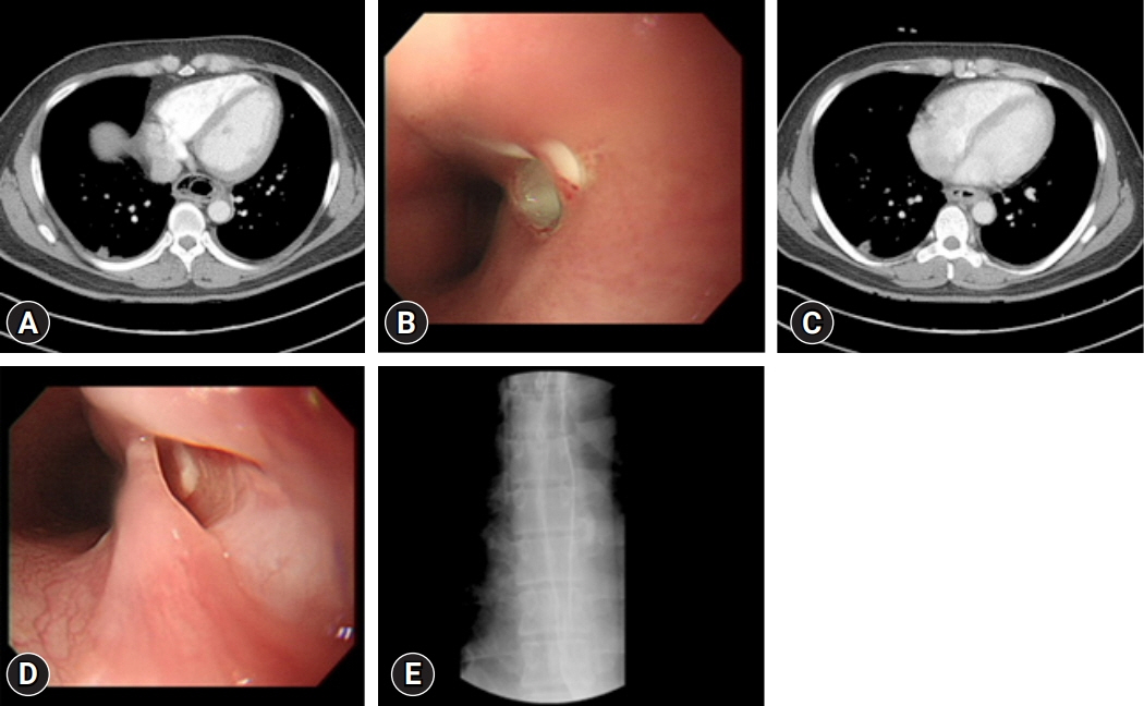
Fig. 1. (A) Enhanced chest computed tomography (CT) shows circumferential wall thickening of the esophagus with multiple air bubbles. (B) Esophagogastroduodenoscopy (EGD) reveals esophageal mucosal edematous change and an upper opening. (C) On hospital day 5, chest CT reveals improvements in esophageal wall thickening, submucosal air, and fluid collection. (D) The previous opening appears as a pseudolumen on follow-up EGD. (E) Esophagogram reveals no passage disturbance or leakage.
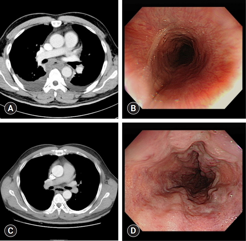
Fig. 2. (A) Circumferential esophageal wall thickening and submucosal fluid collection with pleural effusion on initial chest computed tomography (CT) image. (B) Edematous esophageal mucosa on initial esophagogastroduodenoscopy (EGD) image. (C) Chest CT on hospital day (HD) 18. Improved esophageal wall edema and submucosal fluid collection. (D) EGD on HD 12. Esophageal wall edema is improved, and esophageal varices are seen on follow-up EGD.
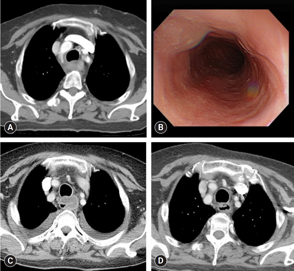
Fig. 3. (A) Diffuse edematous esophageal wall thickening on the initial chest computed tomography (CT). (B) Edematous esophageal wall on initial esophagogastroduodenoscopy. (C) Chest CT on hospital day (HD) 5 reveals increased edema and submucosal fluid collection in the esophageal wall compared with the previous CT. (D) Chest CT on HD 29 reveals decreased esophageal wall thickening and submucosal fluid collection compared with the previous CT.
Reference
-
References
1. Kim HS, Hwang JH, Hong SS, Chang WH, Kim HJ, Chang YW, et al. Acute diffuse phlegmonous esophagogastritis: a case report. J Korean Med Sci. 2010; 25:1532–5.
Article2. Chang PC, Wang WL, Hwang TZ, Cheng YJ. Intramural dissection with mucosal rupture alleviating phlegmonous esophagitis. Eur J Cardiothorac Surg. 2012; 41:442–4.
Article3. Yun SM, Jeong YJ, Hwang M, Lee G, Lee JW, Kim GH, et al. Usefulness of contrast-enhanced CT in a patient with acute phlegmonous esophagitis: a case report and literature review. Medicina (Kaunas). 2022; 58:864.
Article4. Woo WG, Do YW, Lee GD, Lee SS. phlegmonous esophagitis treated with internal drainage and feeding jejunostomy. Korean J Thorac Cardiovasc Surg. 2017; 50:453–5.
Article5. Kim GY, Ward J, Henessey B, Peji J, Godell C, Desta H, et al. Phlegmonous gastritis: case report and review. Gastrointest Endosc. 2005; 61:168–74.
Article6. Kim JW, Ahn HY, Kim GH, Kim YD, Cho JS. Endoscopic intraluminal drainage: an alternative treatment for phlegmonous esophagitis. Korean J Thorac Cardiovasc Surg. 2019; 52:165–9.
Article7. Hsu CY, Liu JS, Chen DF, Shih CC. Acute diffuse phlegmonous esophagogastritis: report of a survived case. Hepatogastroenterology. 1996; 43:1347–52.8. Yun CH, Cheng SM, Sheu CI, Huang JK. Acute phlegmonous esophagitis: an unusual case (2005: 8b). Eur Radiol. 2005; 15:2380–1.
Article
- Full Text Links
-
- Actions
-
Cited
- CITED
-
- Close
- Share
-
- Similar articles
-
- Phlegmonous Esophagitis Treated with Internal Drainage and Feeding Jejunostomy
- Acute Phlegmonous Esophagitis with Mediastinitis Complicated by an Esophageal Perforation: A Case Report
- Endoscopic Intraluminal Drainage: An Alternative Treatment for Phlegmonous Esophagitis
- A Case of Acute Phlegmonous Esophagitis
- Esophageal Stricture after Endoscopic Drainage of Esophageal Abscess as a Complication of Acute Phlegmonous Esophagitis: A Case Report
