Esophageal Stricture after Endoscopic Drainage of Esophageal Abscess as a Complication of Acute Phlegmonous Esophagitis: A Case Report
- Affiliations
-
- 1Department of Internal Medicine, Pusan National University Yangsan Hospital, Yangsan, Korea
- KMID: 2537097
- DOI: http://doi.org/10.4166/kjg.2022.098
Abstract
- Esophageal abscess caused by acute phlegmonous esophagitis is rare but life-threatening. Rapid abscess drainage is an important part of the treatment, and endoscope-assisted intra-luminal abscess drainage is frequently performed. Although endoscopic drainage is less invasive than surgery, it has the potential to cause esophageal stricture as a complication. We present a rare case of esophageal stricture as a complication of intra-luminal drainage and evaluate a method to minimize the incidence of esophageal stricture complications.
Keyword
Figure
-
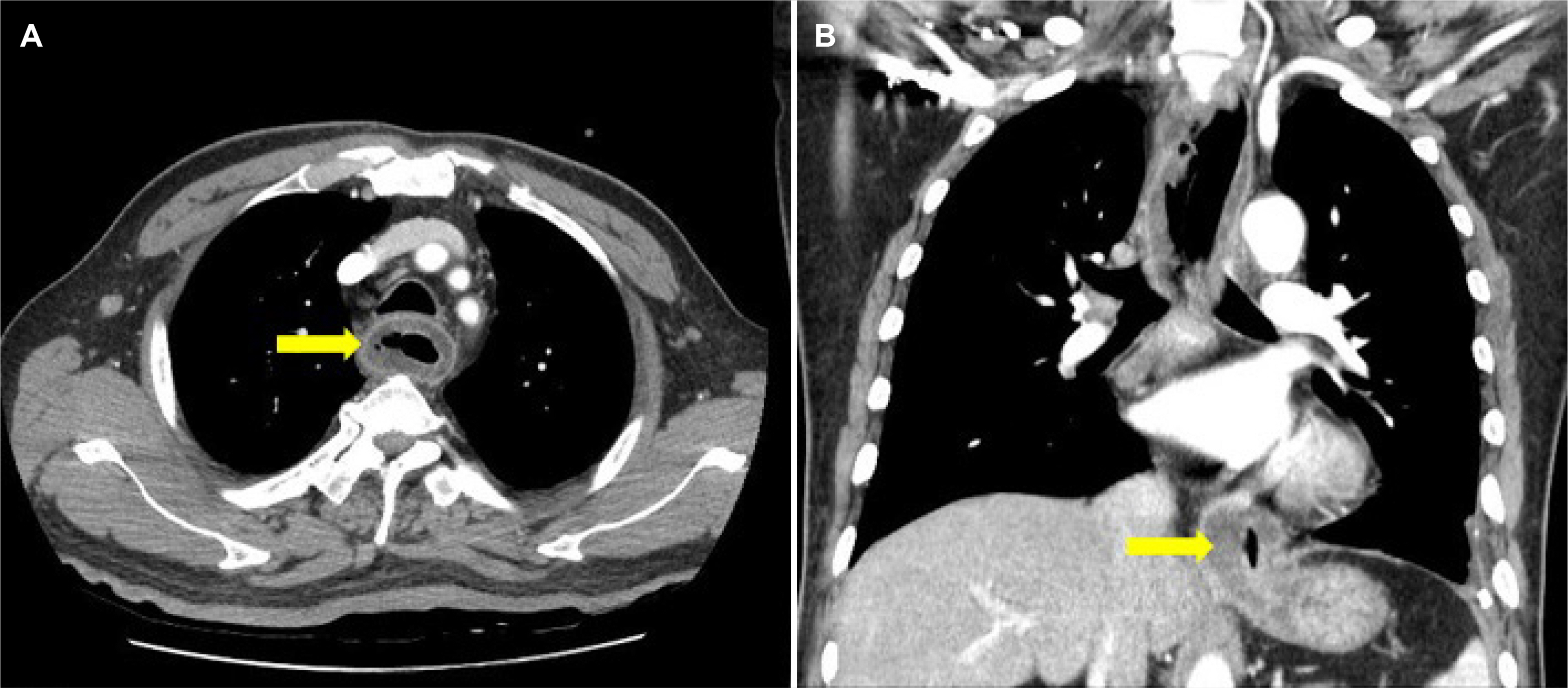
Fig. 1 Initial chest CT image. (A) Diffuse wall thickening of the esophageal wall and air-bubble sign (arrow) were observed. (B) Esophageal dilatation due to the mass like lesion (arrow) of the gastroesophageal junction is observed.
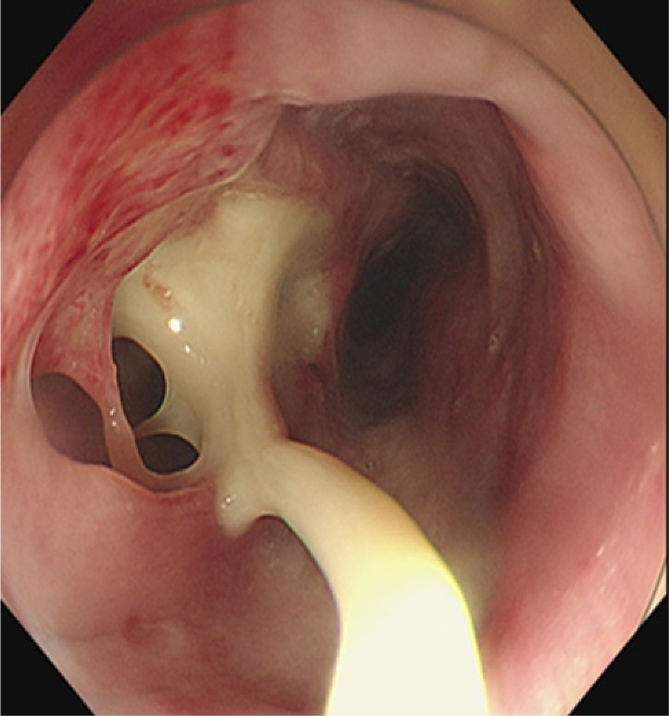
Fig. 2 Initial gastric endoscopy image. An abscess of from 23 cm to 45 cm of the incisor teeth accompanied with 2 cm sized mucosal defect on incisor teeth 23 cm of esophageal wall.

Fig. 3 Endoscopic drainage of an abscess in the esophagus. (A) Abscess lumen (arrow) is observed. (B) Esophageal mucosal incision was made from the incisor teeth 23 cm to 40 cm.

Fig. 4 Esophageal stricture (arrow) was newly revealed following endoscopy.
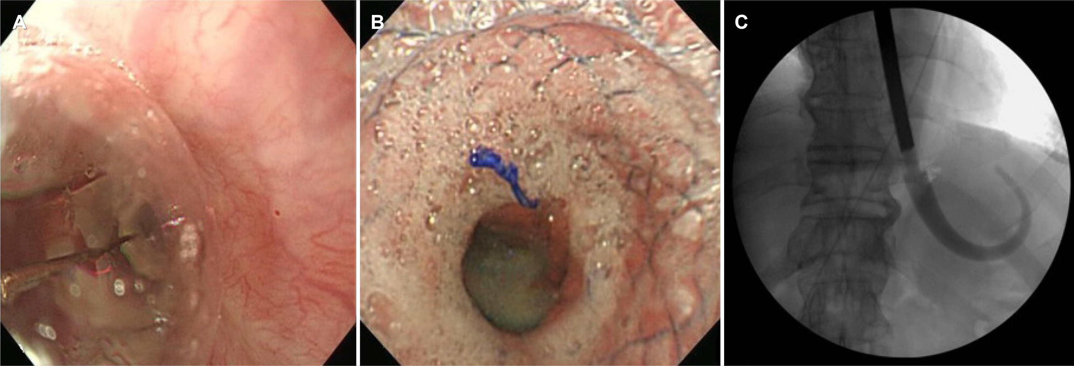
Fig. 5 Treatment for esophageal stricture. (A) Endoscopic balloon dilation. (B) Esophageal self-expandable metal stent insertion. (C) Esophageal lumen dilatation using the bougienage.
Reference
-
1. Muir AD, White J, McGuigan JA, McManus KG, Graham AN. 2003; Treatment and outcomes of oesophageal perforation in a tertiary referral centre. Eur J Cardiothorac Surg. 23:799–804. DOI: 10.1016/S1010-7940(03)00050-2. PMID: 12754036.
Article2. Tonouchi A, Kuwabara S, Furukawa K, Matsuzawa N, Kobayashi K. 2017; Phlegmonous esophagitis treated by endoscopic drainage. Esophagus. 14:183–187. DOI: 10.1007/s10388-016-0562-4.
Article3. Chang PC, Wang WL, Hwang TZ, Cheng YJ. 2012; Intramural dissection with mucosal rupture alleviating phlegmonous esophagitis. Eur J Cardiothorac Surg. 41:442–444. DOI: 10.1016/j.ejcts.2011.06.027. PMID: 21807529.
Article4. Kim HS, Hwang JH, Hong SS, et al. 2010; Acute diffuse phlegmonous esophagogastritis: a case report. J Korean Med Sci. 25:1532–1535. DOI: 10.3346/jkms.2010.25.10.1532. PMID: 20890440. PMCID: PMC2946669.
Article5. Kim JW, Ahn HY, Kim GH, Kim YD, Hoseok , Cho JS. 2019; Endoscopic intraluminal drainage: an alternative treatment for phlegmonous esophagitis. Korean J Thorac Cardiovasc Surg. 52:165–169. DOI: 10.5090/kjtcs.2019.52.3.165. PMID: 31236377. PMCID: PMC6559182.
Article6. Kim JH, Song HY, Kim HC, et al. 2008; Corrosive esophageal strictures: long-term effectiveness of balloon dilation in 117 patients. J Vasc Interv Radiol. 19:736–741. DOI: 10.1016/j.jvir.2008.01.015. PMID: 18440463.
Article7. Ruigómez A, García Rodríguez LA, Wallander MA, Johansson S, Eklund S. 2006; Esophageal stricture: incidence, treatment patterns, and recurrence rate. Am J Gastroenterol. 101:2685–2692. DOI: 10.1111/j.1572-0241.2006.00828.x. PMID: 17227515.
Article8. Pasha SF, Acosta RD, et al. ASGE Standards of Practice Committee. 2014; The role of endoscopy in the evaluation and management of dysphagia. Gastrointest Endosc. 79:191–201. DOI: 10.1016/j.gie.2013.07.042. PMID: 24332405.
Article9. Zhang Y, Zhang B, Wang Y, et al. 2020; Advances in the prevention and treatment of esophageal stricture after endoscopic submucosal dissection of early esophageal cancer. J Transl Int Med. 8:135–145. DOI: 10.2478/jtim-2020-0022. PMID: 33062589. PMCID: PMC7534493.
Article10. Jha S, Levine MS, Rubesin SE, et al. 2006; Detection of strictures on upper gastrointestinal tract radiographic examinations after laparoscopic Roux-en-Y gastric bypass surgery: importance of projection. AJR Am J Roentgenol. 186:1090–1093. DOI: 10.2214/AJR.05.0160. PMID: 16554584.
Article11. Van Boeckel PG, Siersema PD. 2015; Refractory esophageal strictures: what to do when dilation fails. Curr Treat Options Gastroenterol. 13:47–58. DOI: 10.1007/s11938-014-0043-6. PMID: 25647687. PMCID: PMC4328110.
Article12. Dan DT, Gannavarapu B, Lee JG, Chang K, Muthusamy VR. 2014; Removable esophageal stents have poor efficacy for the treatment of refractory benign esophageal strictures (RBES). Dis Esophagus. 27:511–517. DOI: 10.1111/j.1442-2050.2012.01432.x. PMID: 23121426.
Article13. Orive-Calzada A, Bernal-Martinez A, Navajas-Laboa M, et al. 2012; Efficacy of intralesional corticosteroid injection in endoscopic treatment of esophageal strictures. Surg Laparosc Endosc Percutan Tech. 22:518–522. DOI: 10.1097/SLE.0b013e3182747b31. PMID: 23238379.
Article14. Yan X, Nie D, Zhang Y, Chang H, Huang Y. 2019; Effectiveness of an orally administered steroid gel at preventing restenosis after endoscopic balloon dilation of benign esophageal stricture. Medicine (Baltimore). 98:e14565. DOI: 10.1097/MD.0000000000014565. PMID: 30813172. PMCID: PMC6407972.
Article15. Yu M, Tan Y, Liu D. 2019; Strategies to prevent stricture after esophageal endoscopic submucosal dissection. Ann Transl Med. 7:271. DOI: 10.21037/atm.2019.05.45. PMID: 31355238. PMCID: PMC6614329.
Article
- Full Text Links
-
- Actions
-
Cited
- CITED
-
- Close
- Share
-
- Similar articles
-
- Acute Phlegmonous Esophagitis with Mediastinitis Complicated by an Esophageal Perforation: A Case Report
- Phlegmonous Esophagitis Treated with Internal Drainage and Feeding Jejunostomy
- Treatment of Phlegmonous Esophagitis Combined with Mediastinitis
- Endoscopic Intraluminal Drainage: An Alternative Treatment for Phlegmonous Esophagitis
- Acute Esophageal Stricture After Induction Chemotherapy for Acute Leukemi: Report of a case
