Succinate-treated macrophages attenuate dextran sodium sulfate colitis in mice
- Affiliations
-
- 1Department of Internal Medicine and Institute of Gastroenterology, Yonsei University College of Medicine, Seoul, Korea
- 2Brain Korea 21 PLUS Project for Medical Science, Yonsei University College of Medicine, Seoul, Korea
- 3Severance Biomedical Science Institute, Yonsei University College of Medicine, Seoul, Korea
- KMID: 2518691
- DOI: http://doi.org/10.5217/ir.2020.00075
Abstract
- Background/Aims
The safety and effectiveness of adalimumab was demonstrated in a phase 3 trial in Japanese patients with intestinal Behçet’s disease. The aim of this study was to evaluate the long-term safety and effectiveness of adalimumab in Japanese patients with intestinal Behçet’s disease.
Figure
-
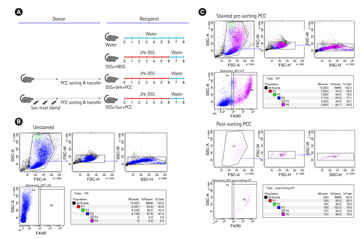
Fig. 1. Peritoneal cavity cell transfer in a dextran sulfate sodium (DSS)-colitis model. (A) Experimental design of the peritoneal cavity cells (PCCs) transfer. Donor mice were intraperitoneally injected with 40 mM succinate or not for 3 days. Recipient mice were transferred with the PCCs on day 0, were then administrated 2% DSS in drinking water until day 6 followed by normal water for 2 days and were sacrificed on day 8. (B, C) Gating strategy of peritoneal macrophage sorting. F4/80+ PCCs isolation using BD FACS Aria II. (B) Unstained PCCs. (C) Stained pre-sorting PCCs and post-sorting PCCs. HBSS, Hanks’ balanced salt solution; Veh, vehicle; Suc, succinate; SSC-A, side-scatter area; FSC-A, forward-scatter area; FSC-W, forward-scatter width; FSC-H, forward-scatter height; SSC-W, side-scatter width; SSC-H, sidescatter height; WT, water.
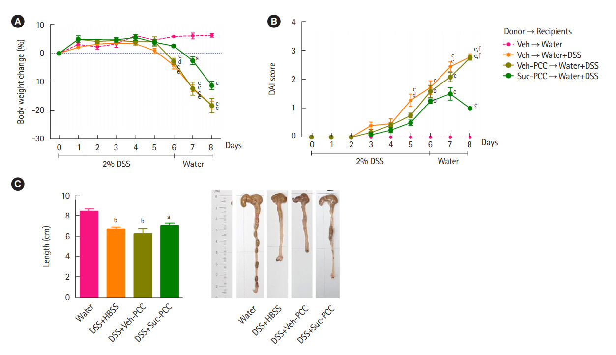
Fig. 2. Transferred succinate-treated peritoneal cavity cells (Suc-PCCs) ameliorate dextran sulfate sodium (DSS)-induced colitis. The recipients of Suc-PCCs show the attenuated body weight change (A), disease activity index (DAI) (B), and colon length (C) compared to vehicle-treated PCC (Veh-PCC). The results were examined using Prism 5.0 software (GraphPad Inc.). Statistical significance was evaluated using one-way analysis of variance and P<0.05 were regarded to be significant. Bars in graphs represent mean±standard error of the mean. aP<0.05, bP<0.01, and cP<0.001 compared with the water group. dP<0.05, eP<0.01, and fP<0.001 compared with the Suc-PCC group. HBSS, Hanks’ balanced salt solution.
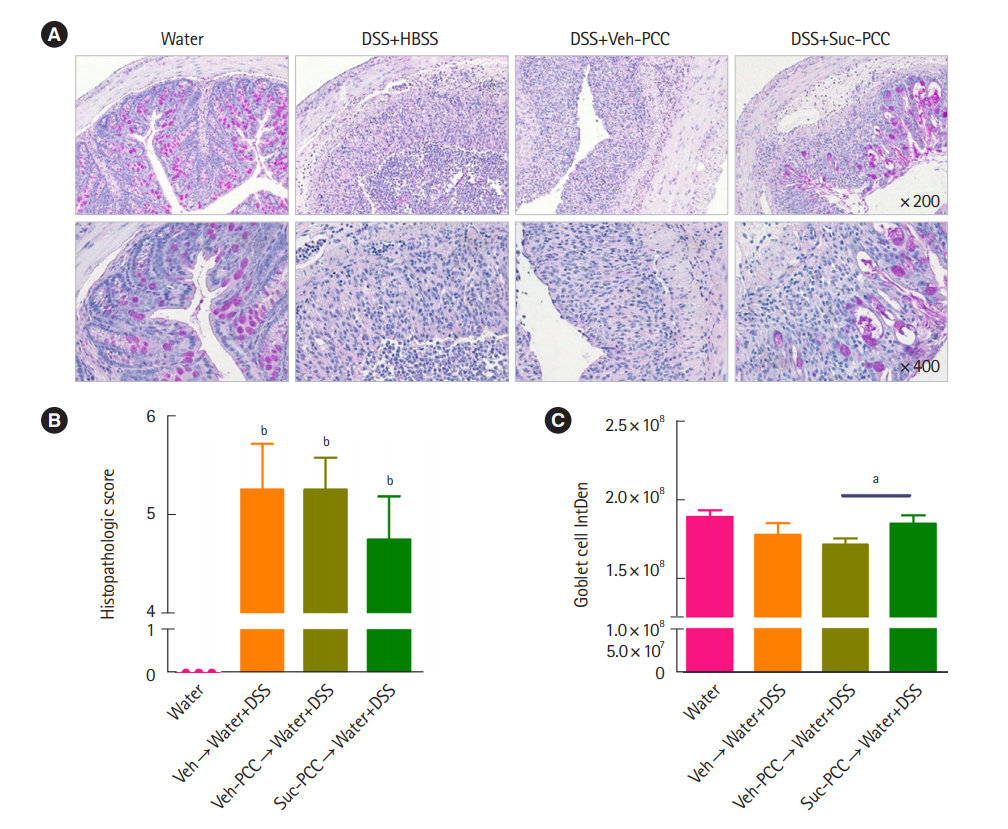
Fig. 3. Periodic acid Schiff staining and quantification of colitis. (A) Section from colon tissues stained with periodic acid Schiff. Histopathologic scores (B) and quantification with integrated density (IntDen) of goblet cells (C) of colon tissues from mice were assessed by Image J v.1.496 software (National Institutes of Health). The results were examined using Prism 5.0 software (GraphPad Inc.). Statistical significance was evaluated using one-way analysis of variance and t-test, respectively, P<0.05 were considered significant. Bars in graphs represent mean±standard error of the mean. aP<0.05, bP<0.001 compared with the water group in (B). DSS, dextran sulfate sodium; HBSS, Hanks’ balanced salt solution; PCC, peritoneal cavity cell; Veh, vehicle; Suc, succinate.
Cited by 2 articles
-
Anti-inflammatory properties of
Escherichia coli Nissle 1917 in a murine colitis model
Jihye Park, Da Hye Kim, Soochan Kim, Hyun Woo Ma, I Seul Park, Mijeong Son, Ji Hyung Kim, Yoojin Shin, Seung Won Kim, Jae Hee Cheon
Intest Res. 2021;19(4):478-481. doi: 10.5217/ir.2021.00121.Anti-inflammatory properties of butyrate-producing atypical
Escherichia coli in a murine colitis model
Ji Hyung Kim, Jee In Yoo, Hyun Woo Ma, I Seul Park, Mijeong Son, Yoojin Shin, Ki Beom Kim, Seung Won Kim, Si Jae Park, Jihye Park
Intest Res. 2023;21(2):266-269. doi: 10.5217/ir.2022.00112.
Reference
-
1. Lloyd-Price J, Arze C, Ananthakrishnan AN, et al. Multi-omics of the gut microbial ecosystem in inflammatory bowel diseases. Nature. 2019; 569:655–662.
Article2. Diskin C, Pålsson-McDermott EM. Metabolic modulation in macrophage effector function. Front Immunol. 2018; 9:270.
Article3. Viola A, Munari F, Sánchez-Rodríguez R, Scolaro T, Castegna A. The metabolic signature of macrophage responses. Front Immunol. 2019; 10:1462.
Article4. Mizoguchi E, Low D, Ezaki Y, Okada T. Recent updates on the basic mechanisms and pathogenesis of inflammatory bowel diseases in experimental animal models. Intest Res. 2020; 18:151–167.
Article5. Macias-Ceja DC, Ortiz-Masiá D, Salvador P, et al. Succinate receptor mediates intestinal inflammation and fibrosis. Mucosal Immunol. 2019; 12:178–187.
Article6. Wang W, Li X, Zheng D, et al. Dynamic changes of peritoneal macrophages and subpopulations during ulcerative colitis to metastasis of colorectal carcinoma in a mouse model. Inflamm Res. 2013; 62:669–680.
Article7. Seo DH, Che X, Kwak MS, et al. Interleukin-33 regulates intestinal inflammation by modulating macrophages in inflammatory bowel disease. Sci Rep. 2017; 7:851.
Article8. Connors J, Dawe N, Van Limbergen J. The role of succinate in the regulation of intestinal inflammation. Nutrients. 2018; 11:25.
Article9. Wu JY, Huang TW, Hsieh YT, et al. Cancer-derived succinate promotes macrophage polarization and cancer metastasis via succinate receptor. Mol Cell. 2020; 77:213–227.
Article10. Keiran N, Ceperuelo-Mallafré V, Calvo E, et al. SUCNR1 controls an anti-inflammatory program in macrophages to regulate the metabolic response to obesity. Nat Immunol. 2019; 20:581–592.
Article
- Full Text Links
-
- Actions
-
Cited
- CITED
-
- Close
- Share
-
- Similar articles
-
- Th17 Responses Are Not Induced in Dextran Sodium Sulfate Model of Acute Colitis
- Histological Study of Experimental Colitis Induced by Dextran Sulfate Sodium
- Anti-inflammatory effects of mulberry twig extracts on dextran sulfate sodium-induced colitis mouse model
- Fine Structure of Goblet Cell Regeneration on Experimental Colitis Induced by Dextran Sulfate Sodium
- Long-Term Effects of Bone Marrow-Derived Mesenchymal Stem Cells in Dextran Sulfate Sodium-Induced Murine Chronic Colitis
