Intraindividual Comparison between Gadoxetate-Enhanced Magnetic Resonance Imaging and Dynamic Computed Tomography for Characterizing Focal Hepatic Lesions: A Multicenter, Multireader Study
- Affiliations
-
- 1Department of Radiology, Research Institute of Radiological Science, Severance Hospital, Yonsei University College of Medicine, Seoul, Korea. kimnex@yuhs.ac
- 2Department of Radiology, Korea University Guro Hospital, Korea University College of Medicine, Seoul, Korea.
- 3Department of Radiology and Research Institute of Radiology, University of Ulsan College of Medicine, Asan Medical Center, Seoul, Korea.
- 4Department of Radiology, Soonchunhyang University Bucheon Hospital, Bucheon, Korea.
- 5Department of Radiology, Samsung Medical Center, Sungkyunkwan University School of Medicine, Seoul, Korea.
- 6Department of Internal Medicine, Severance Hospital, Yonsei University College of Medicine, Seoul, Korea.
- 7Department of Internal Medicine, University of Ulsan College of Medicine, Asan Medical Center, Seoul, Korea.
- 8Department of Internal Medicine, Soonchunhyang University Bucheon Hospital, Bucheon, Korea.
- 9Department of Internal Medicine, Korea University Guro Hospital, Korea University College of Medicine, Seoul, Korea.
- 10Department of Medicine, Samsung Medical Center, Sungkyunkwan University School of Medicine, Seoul, Korea.
- KMID: 2471674
- DOI: http://doi.org/10.3348/kjr.2019.0363
Abstract
OBJECTIVE
To compare the diagnostic accuracy of dynamic computed tomography (CT) and gadoxetate-enhanced magnetic resonance imaging (MRI) for characterization of hepatic lesions by using the Liver Imaging Reporting and Data System (LI-RADS) in a multicenter, off-site evaluation.
MATERIALS AND METHODS
In this retrospective multicenter study, we evaluated 231 hepatic lesions (114 hepatocellular carcinomas [HCCs], 58 non-HCC malignancies, and 59 benign lesions) confirmed histologically in 217 patients with chronic liver disease who underwent both gadoxetate-enhanced MRI and dynamic CT at one of five tertiary hospitals. Four radiologists at different institutes independently reviewed all MR images first and the CT images 4 weeks later. They evaluated the major and ancillary imaging features and categorized each hepatic lesion according to the LI-RADS v2014. Diagnostic performance was calculated and compared using generalized estimating equations.
RESULTS
MRI showed higher sensitivity and accuracy than CT for diagnosing hepatic malignancies; the pooled sensitivities, specificities, and accuracies for categorizing LR-5/5V/M were 59.0% vs. 72.4% (CT vs. MRI; p < 0.001), 83.5% vs. 83.9% (p = 0.906), and 65.3% vs. 75.3% (p < 0.001), respectively. CT and MRI showed comparable capabilities for differentiating between HCC and other malignancies, with pooled accuracies of 79.9% and 82.4% for categorizing LR-M, respectively (p = 0.139).
CONCLUSION
Gadoxetate-enhanced MRI showed superior accuracy for categorizing LR-5/5V/M in hepatic malignancies in comparison with dynamic CT. Both modalities had comparable accuracies for distinguishing other malignancies from HCC.
Keyword
MeSH Terms
Figure
-
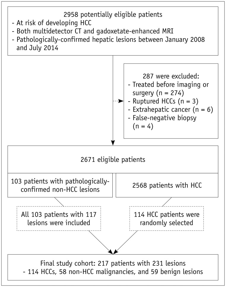
Fig. 1 Flowchart illustrating subject selection.CT = computed tomography, HCC = hepatocellular carcinoma, MRI = magnetic resonance imaging
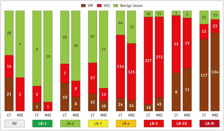
Fig. 2 Frequencies and proportions of HCC, OM, and benign lesions according to imaging modality and Liver Imaging Reporting and Data System category.Numbers and areas of segments in each vertical bar indicate numbers and proportions of HCC, OM, and benign lesions, respectively. Pooled results from four reviewers are shown here (see Supplementary Table 1 for full results). NV = not visible, OM = other malignancies
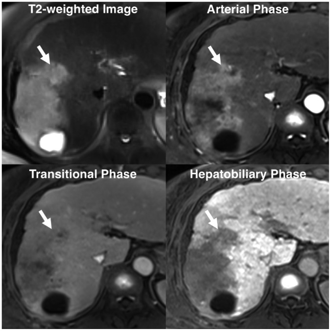
Fig. 3 Non-HCC malignancy with tumor in vein.Diffuse hypervascular tumor with infiltrative margins is seen at right hemi-liver. Tumor also invades adjacent portal vein branch (P8), forming mass within vein (arrows). Two of our four reviewers categorized this mass as LR-5V, while other two assigned score of LR-M. Pathologic diagnosis obtained after biopsy was combined HCC-cholangiocarcinoma.
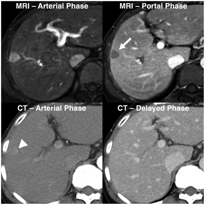
Fig. 4 Pathologically-confirmed HCC categorized as LR-5 only with gadoxetate-enhanced MRI.58-year-old female HBV carrier underwent both dynamic CT and gadoxetate-enhanced MRI with interval of 3 days. Her serum alpha-fetoprotein level was elevated at surveillance for HCC. On gadoxetate-enhanced MRI, all our reviewers found 1.8-cm nodule in right liver (arrow) with arterial hyperenhancement, portal washout appearance, and capsule appearance, and categorized this nodule as LR-5. However, on dynamic CT, only faint arterial hyperenhancement (arrowhead) was visualized at corresponding location. All reviewers categorized lesion as LR-3 or LR-4. HBV = hepatitis B virus
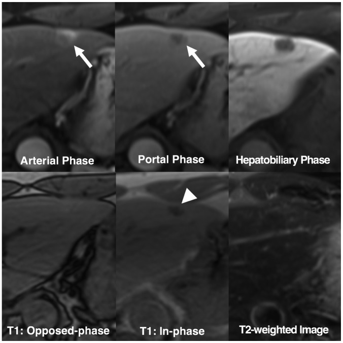
Fig. 5 Benign lesion initially categorized as LR-5 but correctly downgraded by applying ancillary features.52-year-old HBV carrier underwent gadoxetate-enhanced MRI after hepatic nodule was found on ultrasonography. 1.4-cm nodule in left liver shows arterial hyperenhancement (arrow) and washout appearance on portal phase (arrow). Two reviewers considered arterial enhancement as rim-like and categorized nodule as LR-M. Other reviewers initially categorized nodule as LR-5. Nodule shows signal drop from opposed-phase to in-phase of T1-weighted gradient-recalled echo sequence (arrowhead), indicating presence of intralesional iron deposits. Note that nodule shows isointensity on T2-weighted image. These features are uncommon finding in progressed HCCs. After applying these ancillary features, reviewers downgraded their categories to LR-4. This nodule was confirmed as angiomyolipoma after hepatic resection.
Reference
-
1. Marrero JA, Kulik LM, Sirlin CB, Zhu AX, Finn RS, Abecassis MM, et al. Diagnosis, staging, and management of hepatocellular carcinoma: 2018 practice guidance by the American Association for the Study of Liver Diseases. Hepatology. 2018; 68:723–750. PMID: 29624699.
Article2. European Association for the Study of the Liver. EASL clinical practice guidelines: management of hepatocellular carcinoma. J Hepatol. 2018; 69:182–236. PMID: 29628281.3. Omata M, Cheng AL, Kokudo N, Kudo M, Lee JM, Jia J, et al. Asia-Pacific clinical practice guidelines on the management of hepatocellular carcinoma: a 2017 update. Hepatol Int. 2017; 11:317–370. PMID: 28620797.
Article4. Korean Liver Cancer Study Group and Korean National Cancer Center. 2014 Korean Liver Cancer Study Group-National Cancer Center Korea practice guideline for the management of hepatocellular carcinoma. Korean J Radiol. 2015; 16:465–522. PMID: 25995680.5. Kudo M, Trevisani F, Abou-Alfa GK, Rimassa L. Hepatocellular carcinoma: therapeutic guidelines and medical treatment. Liver Cancer. 2016; 6:16–26. PMID: 27995084.
Article6. Kim YY, An C, Kim DY, Aljoqiman KS, Choi JY, Kim MJ. Failure of hepatocellular carcinoma surveillance: inadequate echogenic window and macronodular parenchyma as potential culprits. Ultrasonography. 2019; 38:311–320. PMID: 31079440.
Article7. Rhee H, Kim MJ, Park YN, An C. A proposal of imaging classification of intrahepatic mass-forming cholangiocarcinoma into ductal and parenchymal types: clinicopathologic significance. Eur Radiol. 2019; 29:3111–3121. PMID: 30560357.
Article8. Kim YY, Park MS, Aljoqiman KS, Choi JY, Kim MJ. Gadoxetic acid-enhanced magnetic resonance imaging: hepatocellular carcinoma and mimickers. Clin Mol Hepatol. 2019; 25:223–233. PMID: 30661336.
Article9. Han S, Choi JI, Park MY, Choi MH, Rha SE, Lee YJ. The diagnostic performance of liver MRI without intravenous contrast for detecting hepatocellular carcinoma: a case-controlled feasibility study. Korean J Radiol. 2018; 19:568–577. PMID: 29962863.
Article10. Murakami T, Tsurusaki M. Hypervascular benign and malignant liver tumors that require differentiation from hepatocellular carcinoma: key points of imaging diagnosis. Liver Cancer. 2014; 3:85–96. PMID: 24944999.
Article11. Chernyak V, Fowler KJ, Kamaya A, Kielar AZ, Elsayes KM, Bashir MR, et al. Liver imaging reporting and data system (LI-RADS) version 2018: imaging of hepatocellular carcinoma in at-risk patients. Radiology. 2018; 289:816–830. PMID: 30251931.
Article12. Cerny M, Bergeron C, Billiard JS, Murphy-Lavallée J, Olivié D, Bérubé J, et al. LI-RADS for MR imaging diagnosis of hepatocellular carcinoma: performance of major and ancillary features. Radiology. 2018; 288:118–128. PMID: 29634435.
Article13. Elsayes KM, Kielar AZ, Elmohr MM, Chernyak V, Masch WR, Furlan A, et al. White paper of the Society of Abdominal Radiology hepatocellular carcinoma diagnosis disease-focused panel on LI-RADS v2018 for CT and MRI. Abdom Radiol (NY). 2018; 43:2625–2642. PMID: 30155697.
Article14. Cerny M, Chernyak V, Olivié D, Billiard JS, Murphy-Lavallée J, Kielar AZ, et al. LI-RADS version 2018 ancillary features at MRI. Radiographics. 2018; 38:1973–2001. PMID: 30289735.
Article15. Zech CJ, Ba-Ssalamah A, Berg T, Chandarana H, Chau GY, Grazioli L, et al. Consensus report from the 8th International Forum for Liver Magnetic Resonance Imaging. Eur Radiol. 2019; 8. 05. [Epub ahead of print]. DOI: 10.1007/s00330-019-06369-4.
Article16. Li J, Wang J, Lei L, Yuan G, He S. The diagnostic performance of gadoxetic acid disodium-enhanced magnetic resonance imaging and contrast-enhanced multi-detector computed tomography in detecting hepatocellular carcinoma: a meta-analysis of eight prospective studies. Eur Radiol. 2019; 6. 27. [Epub ahead of print]. DOI: 10.1007/s00330-019-06294-6.
Article17. Korean Society of Abdominal Radiology. Diagnosis of hepatocellular carcinoma with gadoxetic acid-enhanced MRI: 2016 consensus recommendations of the Korean Society of Abdominal Radiology. Korean J Radiol. 2017; 18:427–443. PMID: 28458595.18. Imai Y, Katayama K, Hori M, Yakushijin T, Fujimoto K, Itoh T, et al. Prospective comparison of Gd-EOB-DTPA-enhanced MRI with dynamic CT for detecting recurrence of HCC after radiofrequency ablation. Liver Cancer. 2017; 6:349–359. PMID: 29234638.
Article19. Roberts LR, Sirlin CB, Zaiem F, Almasri J, Prokop LJ, Heimbach JK, et al. Imaging for the diagnosis of hepatocellular carcinoma: a systematic review and meta-analysis. Hepatology. 2018; 67:401–421. PMID: 28859233.
Article20. Hajian-Tilaki K. Sample size estimation in diagnostic test studies of biomedical informatics. J Biomed Inform. 2014; 48:193–204. PMID: 24582925.
Article21. Pencina MJ, D'Agostino RB Sr, D'Agostino RB Jr, Vasan RS. Evaluating the added predictive ability of a new marker: from area under the ROC curve to reclassification and beyond. Stat Med. 2008; 27:157–172. discussion 207-212. PMID: 17569110.
Article22. Liu X, Jiang H, Chen J, Zhou Y, Huang Z, Song B. Gadoxetic acid disodium-enhanced magnetic resonance imaging outperformed multidetector computed tomography in diagnosing small hepatocellular carcinoma: a meta-analysis. Liver Transpl. 2017; 23:1505–1518. PMID: 28886231.
Article23. Choi SH, Lee SS, Kim SY, Park SH, Park SH, Kim KM, et al. Intrahepatic cholangiocarcinoma in patients with cirrhosis: differentiation from hepatocellular carcinoma by using gadoxetic acid-enhanced MR imaging and dynamic CT. Radiology. 2017; 282:771–781. PMID: 27797675.
Article24. Chen N, Motosugi U, Morisaka H, Ichikawa S, Sano K, Ichikawa T, et al. Added value of a gadoxetic acid-enhanced hepatocyte-phase image to the LI-RADS system for diagnosing hepatocellular carcinoma. Magn Reson Med Sci. 2016; 15:49–59. PMID: 26104079.
Article25. Joo I, Lee JM, Lee DH, Ahn SJ, Lee ES, Han JK. Liver imaging reporting and data system v2014 categorization of hepatocellular carcinoma on gadoxetic acid-enhanced MRI: comparison with multiphasic multidetector computed tomography. J Magn Reson Imaging. 2017; 45:731–740. PMID: 27474328.
Article26. Kim YN, Song JS, Moon WS, Hwang HP, Kim YK. Intra-individual comparison of hepatocellular carcinoma imaging features on contrast-enhanced computed tomography, gadopentetate dimeglumine-enhanced MRI, and gadoxetic acid-enhanced MRI. Acta Radiol. 2018; 59:639–648. PMID: 28825310.
Article27. Fraum TJ, Tsai R, Rohe E, Ludwig DR, Salter A, Nalbantoglu I, et al. Differentiation of hepatocellular carcinoma from other hepatic malignancies in patients at risk: diagnostic performance of the liver imaging reporting and data system version 2014. Radiology. 2018; 286:158–172. PMID: 28853673.28. Kim MJ, Lee S, An C. Problematic lesions in cirrhotic liver mimicking hepatocellular carcinoma. Eur Radiol. 2019; 29:5101–5110. PMID: 30788586.
Article29. Kim YY, Kim MJ, Kim EH, Roh YH, An C. Hepatocellular carcinoma versus other hepatic malignancy in cirrhosis: performance of LI-RADS version 2018. Radiology. 2019; 291:72–80. PMID: 30694166.
Article30. Lee HS, Kim MJ, An C. How to utilize LR-M features of the LI-RADS to improve the diagnosis of combined hepatocellular-cholangiocarcinoma on gadoxetate-enhanced MRI? Eur Radiol. 2019; 29:2408–2416. PMID: 30552477.
Article31. Ludwig DR, Fraum TJ, Cannella R, Ballard DH, Tsai R, Naeem M, et al. Hepatocellular carcinoma (HCC) versus non-HCC: accuracy and reliability of liver imaging reporting and data system v2018. Abdom Radiol (NY). 2019; 44:2116–2132. PMID: 30798397.
Article32. Ronot M, Fouque O, Esvan M, Lebigot J, Aubé C, Vilgrain V. Comparison of the accuracy of AASLD and LI-RADS criteria for the non-invasive diagnosis of HCC smaller than 3 cm. J Hepatol. 2018; 68:715–723. PMID: 29274407.33. Becker AS, Barth BK, Marquez PH, Donati OF, Ulbrich EJ, Karlo C, et al. Increased interreader agreement in diagnosis of hepatocellular carcinoma using an adapted LI-RADS algorithm. Eur J Radiol. 2017; 86:33–40. PMID: 28027763.
Article34. Barth BK, Donati OF, Fischer MA, Ulbrich EJ, Karlo CA, Becker A, et al. Reliability, validity, and reader acceptance of LI-RADS— An in-depth analysis. Acad Radiol. 2016; 23:1145–1153. PMID: 27174029.35. Fowler KJ, Tang A, Santillan C, Bhargavan-Chatfield M, Heiken J, Jha RC, et al. Interreader reliability of LI-RADS version 2014 algorithm and imaging features for diagnosis of hepatocellular carcinoma: a large international multireader study. Radiology. 2018; 286:173–185. PMID: 29091751.
Article
- Full Text Links
-
- Actions
-
Cited
- CITED
-
- Close
- Share
-
- Similar articles
-
- A Comprehensive Review of Hepatocellular Carcinoma Enhancement Patterns in MRI: Emphasis on Gadoxetate-Enhanced Imaging
- Hemangioma Diagnosed by Gadoxetate Disodium-Enhanced MRI in a Patient with Chronic Hepatitis C
- Fusion imaging of real-time ultrasonography with CT or MRI for hepatic intervention
- Hepatic Arterioportal Shunts: Dynamic CT and MR Features
- Hepatic Angiomyolipoma Presenting as a Hyperintense Lesion During the Hepatobiliary Phase of Gadoxetic Acid Enhanced-MRI: a Case Report
