Diagnostic Utility of Polymerase Chain Reaction for Acanthamoeba in Contact Lens-related Keratitis with Epithelial Defects
- Affiliations
-
- 1Department of Ophthalmology, Samsung Medical Center, Sungkyunkwan University School of Medicine, Seoul, Korea. ldhlse@gmail.com
- 2Department of Laboratory Medicine and Genetics, Samsung Medical Center, Sungkyunkwan University School of Medicine, Seoul, Korea.
- KMID: 2466188
- DOI: http://doi.org/10.3341/jkos.2019.60.12.1312
Abstract
- PURPOSE
To report contact lens related Acanthamoeba keratitis with corneal epithelial defect cases which were diagnosed using polymerase chain reaction (PCR).
CASE SUMMARY
A 51-year-old male visited our hospital for loss of visual acuity and ocular pain in both eyes. He had been wearing therapeutic contact lenses in both eyes for 4 days prior to his visit, and showed a corneal epithelial defect with corneal edema in both eyes. The corneal edema did not improve after treatment for bacterial and herpes keratitis, so we conducted PCR for Acanthamoeba using the aqueous fluid in the anterior chamber, which showed positive results. A 32-year-old male complained of low visual acuity and ocular pain in both eyes. He had a history of corneal erosion. He had been wearing therapeutic contact lenses in both eyes for 3 days prior to his visit for a corneal epithelial defect. We suspected recurrent corneal erosion syndrome, but PCR for Acanthamoeba of the corneal scraping showed positive results. A 26-year-old female visited our hospital for ocular pain, and discomfort in her left eye. Because of severe dry eye, she had been wearing therapeutic contact lenses for 6 weeks prior to her visit. Her left eye showed corneal infiltration and epithelial defects. The left cornea scraping was positive for bacteria, and PCR for Acanthamoeba also showed positive results.
CONCLUSIONS
Clinicians should consider the use of PCR for the early diagnosis of Acanthamoeba keratitis in contact lens-related keratitis with corneal epithelial defects.
MeSH Terms
Figure
-
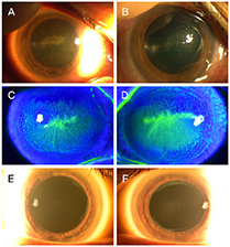
Figure 1 Slit-lamp photograph of the cornea of 51-year-old man with history of wearing therapeutic contacts lenses. (A) Right cornea, (B) left cornea. Central epithelial defect with diffuse corneal edema was seen on both cornea. Slit-lamp photograph with fluorescein stain of the cornea after 2 days. (C) Right cornea, (D) left cornea. Epithelial defect almost healed, dendrite-like lesions remained. Slit-lamp photograph of the cornea after 4 months of treatment covering Acanthamoeba keratitis. (E) Right cornea, (F) left cornea.
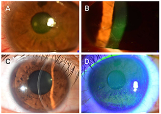
Figure 2 Slit-lamp photograph of the cornea with fluorescein stain of 32-year-old man with history of RCES, and wearing therapeutic contact lenses. (A) Right cornea, (B) left cornea. Corneal epithelial defect was seen on both cornea. Slit lamp photograph after 2 days of treatment covering both RCES and Acanthamoeba keratitis. (C) Right cornea, (D) left cornea. RCES = recurrent corneal erosion syndrome.
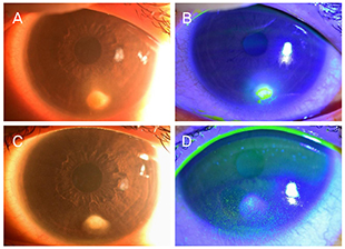
Figure 3 Slit-lamp photograph of the left cornea of 26-year-old woman with history of wearing therapeutic contact lenses. (A) Infiltration with diffuse haze was seen on the left cornea. (B) Fluorescein stained cornea showed epithelial defect in the center of infiltration. (C) Slit-lamp photograph with fluorescein stain of the left cornea. Corneal infiltration improved, but corneal opacity remained. (D) Epithelial defect of cornea showed much improvement.
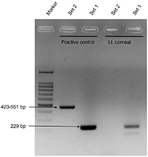
Figure 4 2% Agarose gel electrophoresis of PCR products of amplified 18S rRNA gene from the patient's left corneal swab. (case 3) Marker is a 100bp DNA Ladder. Purified DNA of Acanthamoeba castellanii from Amplirun DNA control (Vircell, Granada, Spain) was used as a positive control. The PCR products using Nelson primer PCR (set 1) and JDP primer PCR (set 2) showed 229 bp and 423 to 551 bp long, respectively. PCR = polymerase chain reaction.
Reference
-
1. Moore MB, McCulley JP, Newton C, et al. Acanthamoeba keratitis. A growing problem in soft and hard contact lens wearers. Ophthalmology. 1987; 94:1654–1661.2. Carnt N, Samarawickrama C, White A, Stapleton F. The diagnosis and management of contact lens-related microbial keratitis. Clin Exp Optom. 2017; 100:482–493.3. Miller DD, Hasan SA, Simmons NL, Stewart MW. Recurrent corneal erosion: a comprehensive review. Clin Ophthalmol. 2019; 13:325–335.4. Bacon AS, Dart JK, Ficker LA, et al. Acanthamoeba keratitis. The value of early diagnosis. Ophthalmology. 1993; 100:1238–1243.5. Kong HH, Shin JY, Yu HS, et al. Mitochondrial DNA restriction fragment length polymorphism (RFLP) and 18S small-subunit ribosomal DNA PCR-RFLP analyses of Acanthamoeba isolated from contact lens storage cases of residents in southwestern Korea. J Clin Microbiol. 2002; 40:1199–1206.6. Inoue T, Ohashi Y. Utility of real-time PCR analysis for appropriate diagnosis for keratitis. Cornea. 2013; 32 Suppl 1:S71–S76.7. Gatti S, Cevini C, Bruno A, et al. In vitro effectiveness of povidone-iodine on Acanthamoeba isolates from human cornea. Antimicrob Agents Chemother. 1998; 42:2232–2234.8. Naginton J, Watson PG, Playfair TJ, et al. Amoebic infection of the eye. Lancet. 1974; 2:1537–1540.9. Bacon AS, Frazer DG, Dart JK, et al. A review of 72 consecutive cases of Acanthamoeba keratitis, 1984–1992. Eye (Lond). 1993; 7(Pt 6):719–725.10. Feist RM, Sugar J, Tessler H. Radial keratoneuritis in Pseudomonas keratitis. Arch Ophthalmol. 1991; 109:774–775.11. Szentmary N, Daas L, Shi L, et al. Acanthamoeba keratitis - clinical signs, differential diagnosis and treatment. J Curr Ophthalmol. 2019; 31:16–23.12. Pfister DR, Cameron JD, Krachmer JH, Holland EJ. Confocal microscopy findings of Acanthamoeba keratitis. Am J Ophthalmol. 1996; 121:119–128.13. Boggild AK, Martin DS, Lee TY, et al. Laboratory diagnosis of amoebic keratitis: comparison of four diagnostic methods for different types of clinical specimens. J Clin Microbiol. 2009; 47:1314–1318.14. McClellan K, Coster DJ. Acanthamoebic keratitis diagnosed by paracentesis and biopsy and treated with propamidine. Br J Ophthalmol. 1987; 71:734–736.15. Dang Burgener NP, Baglivo E, Farpour B, et al. Acanthamoeba detection in the anterior chamber. Br J Ophthalmol. 2006; 90:649–650.
- Full Text Links
-
- Actions
-
Cited
- CITED
-
- Close
- Share
-
- Similar articles
-
- Contamination of Acanthamoeba in Contact Lens Care System
- Acanthoamoeba Keratitis Induced by a Therapeutic, Soft Contact Lens: Diagnosis via Gram Staining
- Contamination of Contact Lens or Contact Lens Storage Case in Contact Lens Related Infectious Keratitis
- Acanthamoeba Keratitis Related to Cosmetic Contact Lenses
- Comparison of Usefulness of Laboratory Diagnosis in Ancanthamoeba Keratitis
