Posterior Inferior Cerebellar Artery Infarction Originating at C1-2 after C1-2 Fusion
- Affiliations
-
- 1Department of Neurosurgery, Gyeongsang National University School of Medicine, Jinju, Korea. leeys1026@hanmail.net
- 2Department of Anesthesiology and Pain Medicine, Gyeongsang National University School of Medicine, Jinju, Korea.
- KMID: 2461127
- DOI: http://doi.org/10.13004/kjnt.2019.15.e27
Abstract
- Vertebral artery injuries associated with C1 lateral mass screw insertion rarely occur during C1-2 fusion. The posterior inferior cerebellar artery (PICA) is uncommonly located at the C1 lateral mass insertion position. A 71-year-old woman with atlanto-axial subluxation and cord compression underwent C1-2 fusion. Sixth nerve palsy and diplopia were detected postoperatively, and decreased consciousness occurred on postoperative day 4. Brain magnetic resonance image (MRI) and computed tomography (CT) revealed PICA infarction. In the preoperative CT angiography, the PICA originated between the C1 and C2 level. In the postoperative CT scan, the PICA was not visible. The patient was treated conservatively for two weeks and recovered. PICA originating between the C1 and C2 level comprises 1.1-1.3% of cases. Therefore, vertebral artery anomalies should be evaluated prior to C1-2 fusion to prevent vessel injuries.
Keyword
MeSH Terms
Figure
-
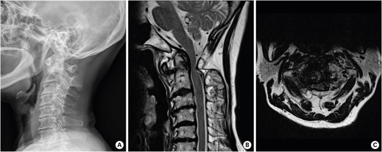
FIGURE 1 Preoperative images. (A) C-spine plain radiography, lateral view (ADI = 5 mm, PADI = 10.4 mm). (B, C) C-spine magnetic resonance image T2 sagittal and T2 axial view showing cord compression with right foraminal stenosis on C1-2.ADI: atlanto-dens interval, PADI: posterior atlanto-dens-interval.
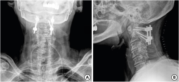
FIGURE 2 Postoperative plain radiographics. (A) Anteroposterior view and (B) Lateral view.
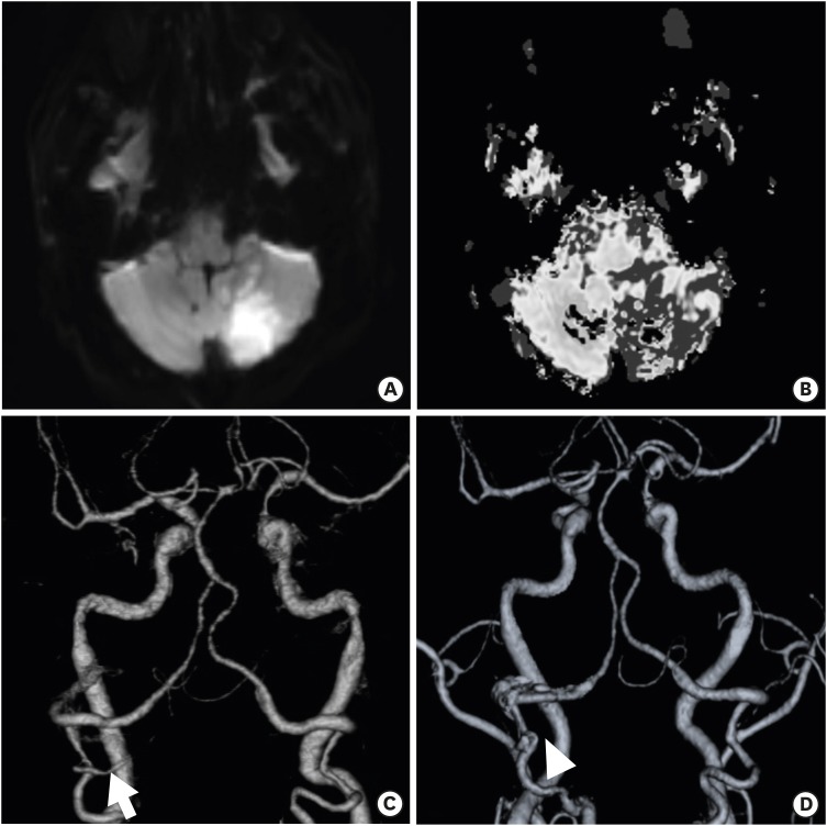
FIGURE 3 Postoperative brain magnetic resonance images and computed tomography angiography images taken on postoperative day 4. (A) Diffusion-weighted image, (B) Mean transit time image showing acute infarction at the left cerebellum, and (C, D) Comparison with preoperative image (arrow). The left posterior inferior cerebellar artery is not shown (arrowhead).
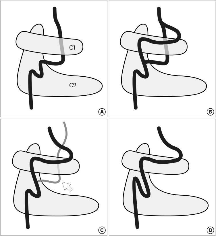
FIGURE 4 Schemas of vertebral artery variation observed at the C1-2 level (left lateral view). (A) Persistent first intersegmental artery, (B) Fenestration of the vertebral artery above and below C1, (C) Posterior inferior cerebellar artery (white arrow) originating from the C1-2 level, and (D) High-riding vertebral artery.
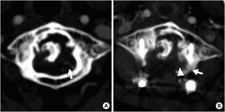
FIGURE 5 Comparisons between preoperative and postoperative images (day 4) of computed tomography angiography. (A) The left posterior inferior cerebellar artery begins between the C1-2 and progresses cephalad towards the cerebellum in the preoperative image (white arrow). (B) The left posterior inferior cerebellar artery may be injured or compressed by the left C1 lateral mass screw (white arrow), and the artery is not seen on the postoperative image (white arrowhead).
Cited by 1 articles
-
Endovascular Management of a Ruptured Aneurysm on a Posterior Inferior Cerebellar Artery with Extradural C2-Origin: Case Report and Literature Review
Rasmus Holmboe Dahl, Gary Lloyd Horn Jr, Zeyad Metwalli, Shankar Prakash Gopinath, Goetz Benndorf
Neurointervention. 2024;19(2):129-134. doi: 10.5469/neuroint.2024.00136.
Reference
-
1. Garcia Alzamora M, Rosahl SK, Lehmberg J, Klisch J. Life-threatening bleeding from a vertebral artery pseudoaneurysm after anterior cervical spine approach: endovascular repair by a triple stent-in-stent method. Case report. Neuroradiology. 47:282–286. 2005; PMID: 15789201.
Article2. Burke JP, Gerszten PC, Welch WC. Iatrogenic vertebral artery injury during anterior cervical spine surgery. Spine J. 5:508–514. 2005; PMID: 16153577.
Article3. Cosgrove GR, Théron J. Vertebral arteriovenous fistula following anterior cervical spine surgery. Report of two cases. J Neurosurg. 66:297–299. 1987; PMID: 3806213.4. Currier BL, Todd LT, Maus TP, Fisher DR, Yaszemski MJ. Anatomic relationship of the internal carotid artery to the C1 vertebra: a case report of cervical reconstruction for chordoma and pilot study to assess the risk of screw fixation of the atlas. Spine. 28:E461–E467. 2003; PMID: 14624095.
Article5. Fine AD, Cardoso A, Rhoton AL Jr. Microsurgical anatomy of the extracranial-extradural origin of the posterior inferior cerebellar artery. J Neurosurg. 91:645–652. 1999; PMID: 10507387.
Article6. Fink KR, Fink JR, Cohen WA. Cervical collaterals may protect against stroke after blunt vertebral artery injury. Emerg Radiol. 18:545–549. 2011; PMID: 21901449.
Article7. Hsu WK, Kannan A, Mai HT, Fehlings MG, Smith ZA, Traynelis VC, et al. Epidemiology and outcomes of vertebral artery injury in 16 582 cervical spine surgery patients: an AOSpine North America multicenter study. Global Spine J. 7:21S–27S. 2017.8. Lee CH, Hong JT, Kang DH, Kim KJ, Kim SW, Kim SW, et al. Epidemiology of iatrogenic vertebral artery injury in cervical spine surgery: 21 multicenter studies. World Neurosurg. 126:e1050–e1054. 2019; PMID: 30878743.
Article9. Lunardini DJ, Eskander MS, Even JL, Dunlap JT, Chen AF, Lee JY, et al. Vertebral artery injuries in cervical spine surgery. Spine J. 14:1520–1525. 2014; PMID: 24411832.
Article10. Madawi AA, Casey AT, Solanki GA, Tuite G, Veres R, Crockard HA. Radiological and anatomical evaluation of the atlantoaxial transarticular screw fixation technique. J Neurosurg. 86:961–968. 1997; PMID: 9171174.
Article11. Nassr AN, Swann PP, Huston J 3rd, Abdelfatah MM, Rose PS, Currier BL. Aberrant posterior inferior cerebellar artery injury with C1 lateral mass screw placement: a case report and review of the literature. Spine J. 14:e7–e14. 2014.
Article12. Neo M, Fujibayashi S, Miyata M, Takemoto M, Nakamura T. Vertebral artery injury during cervical spine surgery: a survey of more than 5600 operations. Spine. 33:779–785. 2008; PMID: 18379405.13. Park HK, Jho HD. The management of vertebral artery injury in anterior cervical spine operation: a systematic review of published cases. Eur Spine J. 21:2475–2485. 2012; PMID: 22790563.
Article14. Rampersaud YR, Moro ER, Neary MA, White K, Lewis SJ, Massicotte EM, et al. Intraoperative adverse events and related postoperative complications in spine surgery: implications for enhancing patient safety founded on evidence-based protocols. Spine. 31:1503–1510. 2006; PMID: 16741462.
Article15. Smith MD, Emery SE, Dudley A, Murray KJ, Leventhal M. Vertebral artery injury during anterior decompression of the cervical spine. A retrospective review of ten patients. J Bone Joint Surg Br. 75:410–415. 1993; PMID: 8496209.
Article16. Uchino A, Saito N, Watadani T, Okada Y, Kozawa E, Nishi N, et al. Vertebral artery variations at the C1-2 level diagnosed by magnetic resonance angiography. Neuroradiology. 54:19–23. 2012; PMID: 21340577.
Article17. Uchino A, Saito N, Ishihara S. Double origin of the posterior inferior cerebellar artery diagnosed by MR angiography: a report of two cases. Neuroradiol J. 28:187–189. 2015; PMID: 25923681.18. Wakao N, Takeuchi M, Nishimura M, Riew KD, Kamiya M, Hirasawa A, et al. Vertebral artery variations and osseous anomaly at the C1-2 level diagnosed by 3D CT angiography in normal subjects. Neuroradiology. 56:843–849. 2014; PMID: 25001076.
Article19. Wright NM, Lauryssen C. American Association of Neurological Surgeons/Congress of Neurological Surgeons. Vertebral artery injury in C1-2 transarticular screw fixation: results of a survey of the AANS/CNS section on disorders of the spine and peripheral nerves. J Neurosurg. 88:634–640. 1998; PMID: 9525707.
Article
- Full Text Links
-
- Actions
-
Cited
- CITED
-
- Close
- Share
-
- Similar articles
-
- C1/2 Transarticular Screw Fixation for Complicated Os Odontoideum: Case Report
- A Case of Sudden Hearing Loss due to Posterior Inferior Cerebellar Artery Infarction
- Spontaneous Anterior Atlas Fracture Following C1 Laminectomy without Fusion: A Case Report
- Dyspnea and Dysphagia after Posterior Atlantoaxial Instrumented Fusion
- Three cases of posterior circulation infarction related with cervical manipulation or trauma
