Propofol promotes osteoclastic bone resorption by increasing DC-STAMP expression
- Affiliations
-
- 1Department of Dental Anesthesia and Pain Medicine, School of Dentistry, Pusan National University, Dental Research Institute, Yangsan, Republic of Korea.
- 2Department of Oral Physiology, School of Dentistry, Pusan National University, Yangsan, Republic of Korea.
- 3Department of Anesthesia and Pain Medicine, School of Medicine, Pusan National University, Yangsan, Republic of Korea. shinsw@pusan.ac.kr
- 4Department of Anesthesia and Pain Medicine, College of Medicine, Kosin University, Busan, Korea.
- KMID: 2430212
- DOI: http://doi.org/10.17245/jdapm.2018.18.6.349
Abstract
- BACKGROUND
Propofol is an intravenous anesthetic which has antioxidant effects due to its similarity in molecular structure to α-tocopherol. It has been reported that α-tocopherol increases osteoclast fusion and bone resorption. Here, we investigated the effects of propofol on signaling pathways of osteoclastogenic gene expression, as well as osteoclastogenesis and bone resorption using bone marrow-derived macrophages (BMMs).
METHODS
BMMs were cultured with macrophage colony-stimulating factor (M-CSF) alone or M-CSF plus receptor activator of nuclear factor kappa B ligand (RANKL) in the presence of propofol (0-50 µM) for 4 days. Mature osteoclasts were stained for tartrate-resistant acid phosphatase (TRAP) and the numbers of TRAP-positive multinucleated osteoclasts were counted. To examine the resorption activities of osteoclasts, a bone resorption assay was performed. To identify the mechanism of action of propofol on the formation of multinucleated osteoclasts, we focused on dendritic cell-specific transmembrane protein (DC-STAMP), a protein essential for pre-osteoclastic cell fusion.
RESULTS
Propofol increased the formation of TRAP-positive multinucleated osteoclasts. In addition, the bone resorption assay revealed that propofol increased the bone resorption area on dentin discs. The mRNA expression of DC-STAMP was upregulated most strongly in the presence of both RANKL and propofol. However, SB203580, a p38 inhibitor, significantly suppressed the propofol/RANKL-induced increase in mRNA expression of DC-STAMP.
CONCLUSION
We have demonstrated that propofol enhances osteoclast differentiation and maturation, and subsequently increases bone resorption. Additionally, we identified the regulatory pathway underlying osteoclast cell-cell fusion, which was enhanced by propofol through p38-mediated DC-STAMP expression.
Keyword
MeSH Terms
-
Acid Phosphatase
Antioxidants
Bone Resorption*
Cell Fusion
Dentin
Gene Expression
Macrophage Colony-Stimulating Factor
Macrophages
Molecular Structure
Osteoclasts*
p38 Mitogen-Activated Protein Kinases
Propofol*
RANK Ligand
RNA, Messenger
Acid Phosphatase
Antioxidants
Macrophage Colony-Stimulating Factor
Propofol
RANK Ligand
RNA, Messenger
p38 Mitogen-Activated Protein Kinases
Figure
-
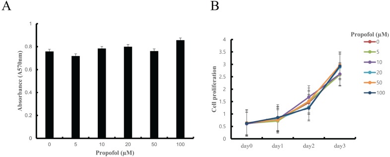
Fig. 1 The effect of propofol on BMM cell viability and proliferation as determined by MTT assay. (A) Propofol did not have a cytotoxic effect on BMMs. (B) Cell proliferation was similar at all concentration of propofol (0–100 µM).
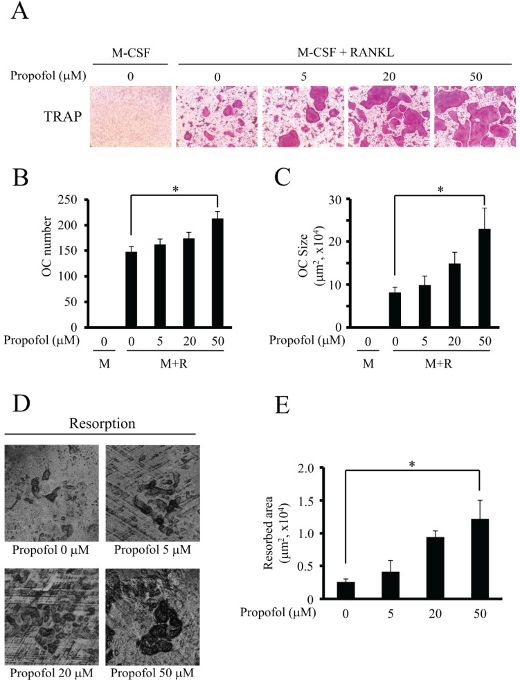
Fig. 2 Propofol markedly augmented the size of osteoclast and subsequently increased bone resorption. (A–C) BMMs were cultured with M-CSF alone or M-CSF plus RANKL with propofol (0–50 µM) for 4 days, and TRAP staining was performed on the resulting osteoclasts. (A) Example images are shown. (B) Quantification of TRAP-positive multinucleated osteoclasts (cells containing more than four nuclei). (C) Quantification of the size of osteoclasts. OC, osteoclast; M, M-CSF; M+R, M-CSF+RANKL. (D–E) To examine the resorption activities of osteoclasts, a bone resorption assay was performed using dentin discs. The resorption pits were visualized and quantified by laser scanning of cell-removed dentin discs. (*, P < 0.05).
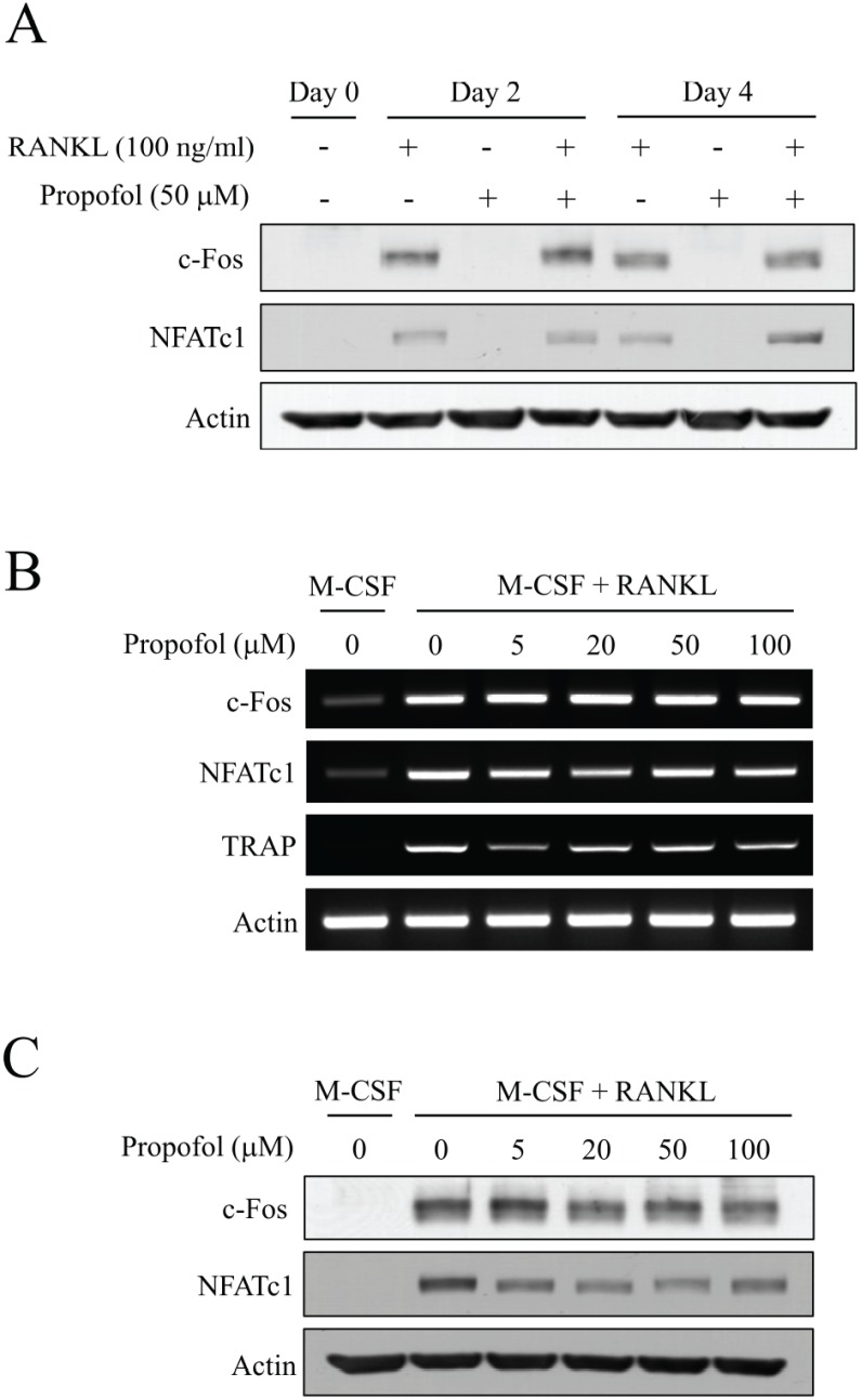
Fig. 3 Propofol has little effect on the expression of c-Fos and NFATc1. (A) BMMs were cultured in the presence or absence of RANKL (100 ng/ml) or propofol (50 mM) for 4 days. Expression of c-Fos and NFATc1 were evaluated by western blotting at day 0, 2 and 4. (B) The mRNA levels of c-Fos and NFATc1 were evaluated by RT-PCR. The mRNA expression of TRAP served as an osteoclastic differentiation marker. (C) The protein expression of c-Fos and NFATc1 at various concentrations of propofol (0–100 µM) was determined by western blotting.
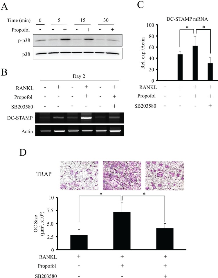
Fig. 4 Propofol increases osteoclast size through the activation of p38 and DC-STAMP expression. (A) Following incubation of BMM cells with propofol for the indicated times, phosphorylation of p38 was evaluated by western blotting. (B) The mRNA expression of DC-STAMP was analyzed by RT-PCR after treatment with the indicated combinations of RANKL (100 ng/ml), propofol (50 µM) and a p38 inhibitor, SB203580 (20 µM) for 2 days. (C) Quantitative real-time PCR analysis of DC-STAMP. (D) The representative TRAP-stained images are shown (top) and the sizes of mature osteoclasts were measured (bottom). Data are mean ± SD (*, P < 0.05). OC, osteoclast.
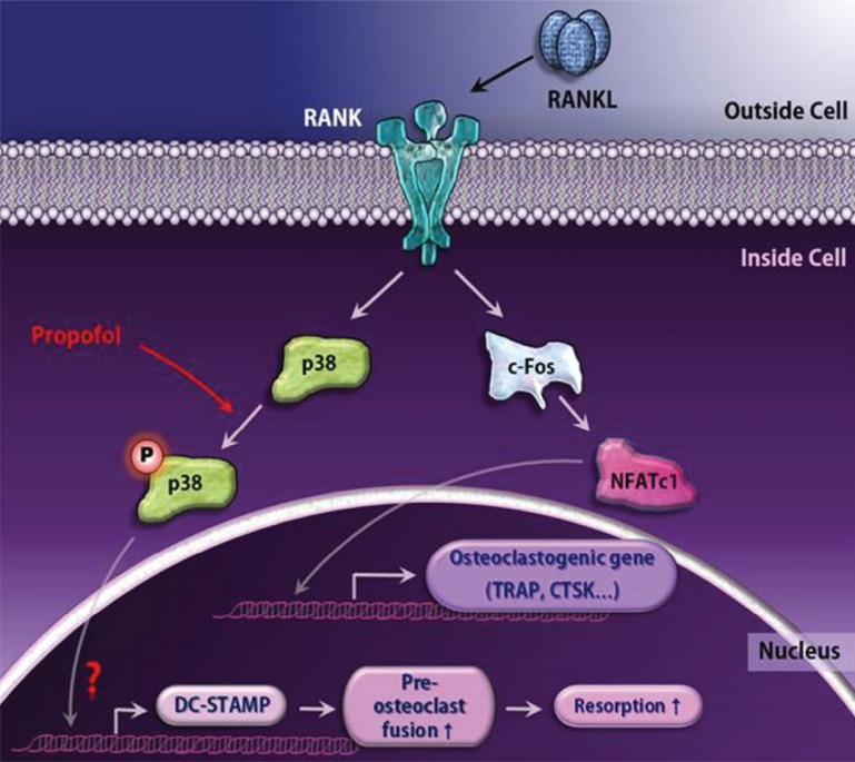
Fig. 5 A schematic diagram for propofol-induced large osteoclast formation. The engagement of RANKL to RANK leads to pivotal osteoclastogenic gene expression via the c-Fos/NFATc1 transcription factor axis. Although propofol does not alter these major differentiation pathways, it functions as a positive regulator of osteoclastogenesis by upregulating phosphorylation of p38 and subsequently increasing DC-STAMP expression.
Reference
-
1. Karsenty G, Kronenberg HM, Settembre C. Genetic control of bone formation. Annu Rev Cell Dev Biol. 2009; 25:629–648. PMID: 19575648.
Article2. Walsh MC, Kim N, Kadono Y, Rho J, Lee SY, Lorenzo J, et al. Osteoimmunology: interplay between the immune system and bone metabolism. Annu Rev Immunol. 2006; 24:33–63. PMID: 16551243.
Article3. Mountzios G, Dimopoulos MA, Bamias A, Papadopoulos G, Kastritis E, Syrigos K, et al. Abnormal bone remodeling process is due to an imbalance in the receptor activator of nuclear factor-kappaB ligand (RANKL)/osteoprotegerin (OPG) axis in patients with solid tumors metastatic to the skeleton. Acta Oncol. 2007; 46:221–229. PMID: 17453373.4. Corral DA, Amling M, Priemel M, Loyer E, Fuchs S, Ducy P, et al. Dissociation between bone resorption and bone formation in osteopenic transgenic mice. Proc Natl Acad Sci USA. 1998; 95:13835–13840. PMID: 9811887.
Article5. Theill LE, Boyle WJ, Penninger JM. RANK-L and RANK: T cells, bone loss, and mammalian evolution. Annu Rev Immunol. 2002; 20:795–823. PMID: 11861618.
Article6. Lacey DL, Timms E, Tan HL, Kelley MJ, Dunstan CR, Burgess T, et al. Osteoprotegerin ligand is a cytokine that regulates osteoclast differentiation and activation. Cell. 1998; 93:165–176. PMID: 9568710.
Article7. Takayanagi H, Kim S, Matsuo K, Suzuki H, Suzuki T, Sato K, et al. RANKL maintains bone homeostasis through c-Fos-dependent induction of interferon-beta. Nature. 2002; 416:744–749. PMID: 11961557.8. Aarts L, van der Hee R, Dekker I, de Jong J, Langemeijer H, Bast A. The widely used anesthetic agent propofol can replace alpha-tocopherol as an antioxidant. FEBS Lett. 1995; 357:83–85. PMID: 8001686.9. Fujita K, Iwasaki M, Ochi H, Fukuda T, Ma C, Miyamoto T, et al. Vitamin E decreases bone mass by stimulating osteoclast fusion. Nat Med. 2012; 18:589–594. PMID: 22388090.
Article10. Hartgers FC, Vissers JL, Looman MW, van Zoelen C, Huffine C, Figdor CG, et al. DC-STAMP, a novel multimembrane-spanning molecule preferentially expressed by dendritic cells. Eur J Immunol. 2000; 30:3585–3590. PMID: 11169400.
Article11. Staege H, Brauchlin A, Schoedon G, Schaffner A. Two novel genes FIND and LIND differentially expressed in deactivated and Listeria-infected human macrophages. Immunogenetics. 2001; 53:105–113. PMID: 11345586.12. Yagi M, Miyamoto T, Sawatani Y, Iwamoto K, Hosogane N, Fujita N, et al. DC-STAMP is essential for cell-cell fusion in osteoclasts and foreign body giant cells. J Exp Med. 2005; 202:345–351. PMID: 16061724.
Article13. Kobayashi N, Kadono Y, Naito A, Matsumoto K, Yamamoto T, Tanaka S, et al. Segregation of TRAF6-mediated signaling pathways clarifies its role in osteoclastogenesis. EMBO J. 2001; 20:1271–1280. PMID: 11250893.
Article14. Gepts E, Camu F, Cockshott ID, Douglas EJ. Disposition of propofol administered as constant rate intravenous infusions in humans. Anesth Analg. 1987; 66:1256–1263. PMID: 3500657.
Article15. Rao A, Luo C, Hogan PG. Transcription factors of the NFAT family: regulation and function. Annu Rev Immunol. 1997; 15:707–747. PMID: 9143705.16. Takayanagi H, Kim S, Koga T, Nishina H, Isshiki M, Yoshida H, et al. Induction and activation of the transcription factor NFATc1 (NFAT2) integrate RANKL signaling in terminal differentiation of osteoclasts. Dev Cell. 2002; 3:889–901. PMID: 12479813.
Article17. Lee ZH, Kim HH. Signal transduction by receptor activator of nuclear factor kappa B in osteoclasts. Biochem Biophys Res Commun. 2003; 305:211–214. PMID: 12745060.
Article18. Teitelbaum SL. RANKing c-Jun in osteoclast development. J Clin Invest. 2004; 114:463–465. PMID: 15314680.
Article19. Matsumoto M, Kogawa M, Wada S, Takayanagi H, Tsujimoto M, Katayama S, et al. Essential role of p38 mitogen-activated protein kinase in cathepsin K gene expression during osteoclastogenesis through association of NFATc1 and PU.1. J Biol Chem. 2004; 279:45969–45979. PMID: 15304486.
Article20. Huang H, Chang EJ, Ryu J, Lee ZH, Lee Y, Kim HH. Induction of c-Fos and NFATc1 during RANKL-stimulated osteoclast differentiation is mediated by the p38 signaling pathway. Biochem Biophys Res Commun. 2006; 351:99–105. PMID: 17052691.
Article21. Luchin A, Purdom G, Murphy K, Clark MY, Angel N, Cassady AI, et al. The microphthalmia transcription factor regulates expression of the tartrate-resistant acid phosphatase gene during terminal differentiation of osteoclasts. J Bone Miner Res. 2000; 15:451–460. PMID: 10750559.
Article22. Mansky KC, Sankar U, Han J, Ostrowski MC. Microphthalmia transcription factor is a target of the p38 MAPK pathway in response to receptor activator of NF-kappa B ligand signaling. J Biol Chem. 2002; 277:11077–11083. PMID: 11792706.23. Boyle WJ, Simonet WS, Lacey DL. Osteoclast differentiation and activation. Nature. 2003; 423:337–342. PMID: 12748652.
Article24. Kim Y, Sato K, Asagiri M, Morita I, Soma K, Takayanagi H. Contribution of nuclear factor of activated T cells c1 to the transcriptional control of immunoreceptor osteoclast-associated receptor but not triggering receptor expressed by myeloid cells-2 during osteoclastogenesis. J Biol Chem. 2005; 280:32905–32913. PMID: 16046394.
Article25. Xing L, Bushnell TP, Carlson L, Tai Z, Tondravi M, Siebenlist U, et al. NF-kappaB p50 and p52 expression is not required for RANK-expressing osteoclast progenitor formation but is essential for RANK- and cytokine-mediated osteoclastogenesis. J Bone Miner Res. 2002; 17:1200–1210. PMID: 12096833.26. Grigoriadis AE, Wang ZQ, Cecchini MG, Hofstetter W, Felix R, Fleisch HA, et al. c-Fos: a key regulator of osteoclast-macrophage lineage determination and bone remodeling. Science. 1994; 266:443–448. PMID: 7939685.
Article27. Sharma SM, Bronisz A, Hu R, Patel K, Mansky KC, Sif S, et al. MITF and PU.1 recruit p38 MAPK and NFATc1 to target genes during osteoclast differentiation. J Biol Chem. 2007; 282:15921–15929. PMID: 17403683.
Article28. Gohda J, Akiyama T, Koga T, Takayanagi H, Tanaka S, Inoue J. RANK-mediated amplification of TRAF6 signaling leads to NFATc1 induction during osteoclastogenesis. EMBO J. 2005; 24:790–799. PMID: 15678102.
Article29. Takayanagi H. Mechanistic insight into osteoclast differentiation in osteoimmunology. J Mol Med (Berl). 2005; 83:170–179. PMID: 15776286.
Article30. Ishii M, Saeki Y. Osteoclast cell fusion: mechanisms and molecules. Mod Rheumatol. 2008; 18:220–227. PMID: 18425565.
Article31. Yagi M, Ninomiya K, Fujita N, Suzuki T, Iwasaki R, Morita K, et al. Induction of DC-STAMP by alternative activation and downstream signaling mechanisms. J Bone Miner Res. 2007; 22:992–1001. PMID: 17402846.
Article
- Full Text Links
-
- Actions
-
Cited
- CITED
-
- Close
- Share
-
- Similar articles
-
- The Effect of Substance P on Osteoclastogenesis and Osteoclastic Bone Resorption in vitro
- Biological characteristics of osteoporosis drugs: the effect of osteoblast–osteoclast coupling
- Regulation of Osteoclasts via Osteoblasts by Alendronate
- The effect of NaF on bone and tooth resorption around an anchor tooth during a rapid maxillary expansion procedure
- Effects of sodium nitroprusside on the formation and activation of the osteoclast in culture
