Spinal Enumeration by Morphologic Analysis of Spinal Variants: Comparison to Counting in a Cranial-To-Caudal Manner
- Affiliations
-
- 1Department of Radiology, Kosin University Gospel Hospital, Busan 49267, Korea. cordialsk@kosin.ac.kr
- KMID: 2424853
- DOI: http://doi.org/10.3348/kjr.2018.19.6.1140
Abstract
OBJECTIVE
To compare the spinal enumeration methods that establish the first lumbar vertebra in patients with spinal variants.
MATERIALS AND METHODS
Of the 1446 consecutive patients who had undergone computed tomography of the spine from March 2012 to July 2016, 100 patients (62 men, 38 women; mean age, 47.9 years; age range, 19-88 years) with spinal variants were included. Two radiologists (readers 1 and 2) established the first lumbar vertebra through morphologic analysis of the thoracolumbar junction, and labeled the vertebra by counting in a cranial-to-caudal manner. Inter-observer agreement was established. Additionally, reader 1 detected the 20th vertebra under the assumption that there are 12 thoracic vertebra, and then classified it as a thoracic vertebra, lumbar vertebra, or thoracolumbar transitional vertebra (TLTV), on the basis of morphologic analysis.
RESULTS
The first lumbar vertebra, as established by morphologic analysis, was labeled by each reader as the 21st segment in 65.0% of the patients, as the 20th segment in 31.0%, and as the 19th segment in 4.0%. Inter-observer agreement between the two readers in determining the first lumbar vertebra, based on morphologic analysis, was nearly perfect (κ value: 1.00). The 20th vertebra was morphologically classified as a TLTV in 60.0% of the patients, as the first lumbar segment in 31.0%, as the second lumbar segment in 4.0%, and as a thoracic segment in 5.0%.
CONCLUSION
The establishment of the first lumbar vertebra using morphologic characteristics of the thoracolumbar junction in patients with spinal variants was consistent with the morphologic traits of vertebral segmentation.
Keyword
MeSH Terms
Figure
-
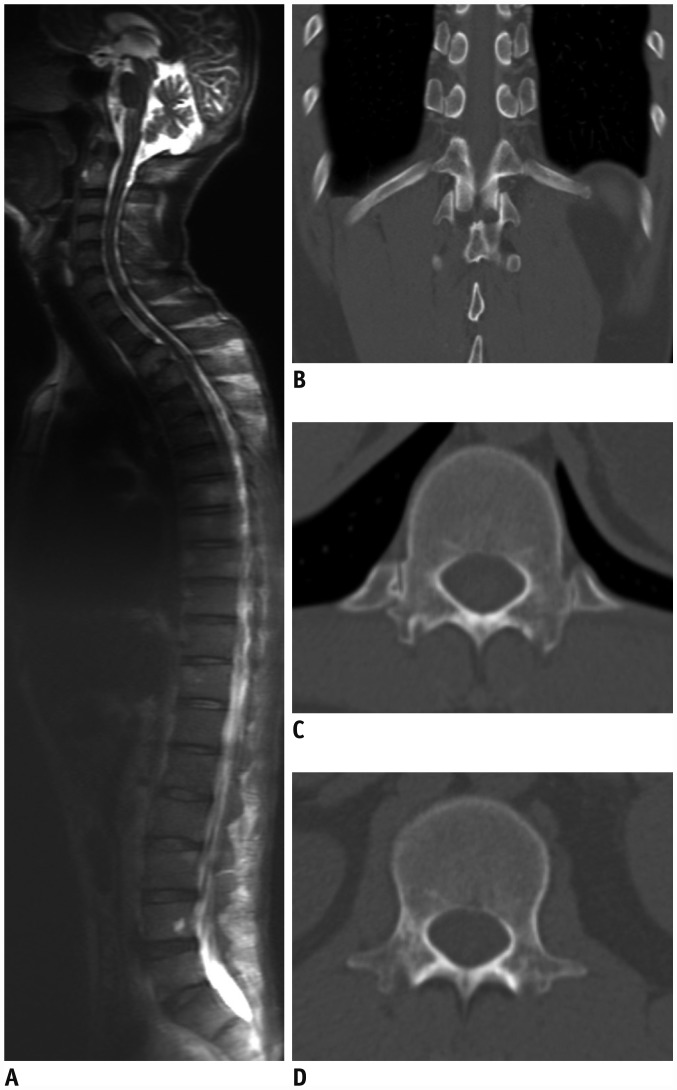
Fig. 1 47-year-old woman with anomalous total number of vertebra.A. Sagittal T2-weighted fast spin-echo magnetic resonance image of whole spine shows 25 presacral mobile vertebra. B, C. Coronal CT image (B) showing 20th vertebra, with paired ribs that are 3.8 cm or greater in length. Paired ribs originate from facet at pedicle of 20th vertebra on axial CT image (C). Therefore, 20th vertebra is morphologically thoracic vertebra. D. On axial CT image, 21st vertebra morphologically appears to be first lumbar vertebra. Thus, this patient has 25 presacral mobile vertebra with seven cervical, 13 thoracic, and five lumbar vertebra. CT = computed tomography
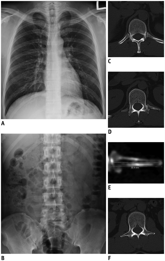
Fig. 2 38-year-old man with TLTV.A, B. Posteroanterior chest radiograph (A) and supine abdominal radiograph (B) demonstrate 24 presacral mobile vertebra. C. Axial CT image showing 19th vertebra, with paired ribs that are 3.8 cm or greater in length and originate from facet at pedicle. Therefore, 19th vertebra is morphologically thoracic vertebra. D. On axial CT image, 20th vertebra, which has short rib on left side and accessory ossification center on right side, is TLTV. E. On curved planar reformatted image of left rib of 20th vertebra, rib length is measured by drawing line at midpoint of rib width from proximal head of rib to distal body. Rib measures 30.4 mm and is classified as short rib. F. On axial CT image, 21st vertebra, exhibiting both fused transverse processes without articulating ribs, is identified as first lumbar vertebra. Thus, this patient has 24 presacral mobile vertebra with seven cervical, 12 thoracic, one TLTV, and four lumbar vertebra. TLTV = thoracolumbar transitional vertebra
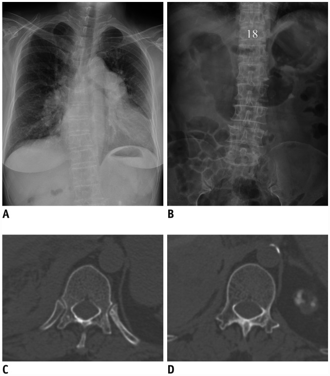
Fig. 3 74-year-old woman with anomalous distribution of vertebra.A, B. Posteroanterior chest radiograph (A) and supine abdominal radiograph (B) indicate presence of 24 presacral mobile vertebra. C. Axial CT image showing 18th vertebra, with paired ribs that are 3.8 cm or greater in length and originate from facet at pedicle. Therefore, 18th vertebra is morphologically thoracic vertebra. D. On axial CT image, 19th vertebra has both fused transverse processes and morphologically appears to be first lumbar vertebra. Thus, this patient has anomalous distribution of 24 presacral mobile vertebra with seven cervical, 11 thoracic, and six lumbar vertebra.
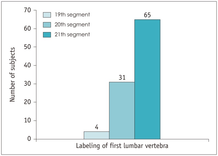
Fig. 4 Labeling of morphologic first lumbar vertebra by counting in cranial-to-caudal manner.
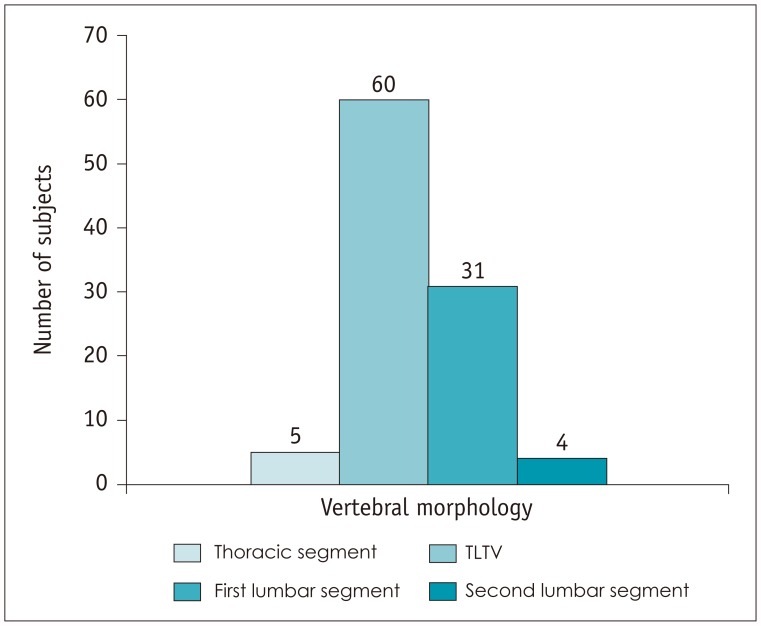
Fig. 5 Morphologic analysis of 20th vertebra.
Cited by 1 articles
-
RE: Spinal Enumeration by Morphologic Analysis of Spinal Variants: Comparison to Counting in a Cranial-To-Caudal Manner
Min Jeong Choi, Sang Yoon Kim
Korean J Radiol. 2019;20(4):693-694. doi: 10.3348/kjr.2018.0834.
Reference
-
1. Narita Y, Kuratani S. Evolution of the vertebral formulae in mammals: a perspective on developmental constraints. J Exp Zool B Mol Dev Evol. 2005; 304:91–106. PMID: 15660398.
Article2. Hanson EH, Mishra RK, Chang DS, Perkins TG, Bonifield DR, Tandy RD, et al. Sagittal whole-spine magnetic resonance imaging in 750 consecutive outpatients: accurate determination of the number of lumbar vertebral bodies. J Neurosurg Spine. 2010; 12:47–55. PMID: 20043764.
Article3. Galis F. Why do almost all mammals have seven cervical vertebrae? Developmental constraints, hox genes, and cancer? J Exp Zool. 1999; 285:19–26. PMID: 10327647.4. Akbar JJ, Weiss KL, Saafir MA, Weiss JL. Rapid MRI detection of vertebral numeric variation. AJR Am J Roentgenol. 2010; 195:465–466. PMID: 20651206.
Article5. Thawait GK, Chhabra A, Carrino JA. Spine segmentation and enumeration and normal variants. Radiol Clin North Am. 2012; 50:587–598. PMID: 22643386.
Article6. Carrino JA, Campbell PD Jr, Lin DC, Morrison WB, Schweitzer ME, Flanders AE, et al. Effect of spinal segment variants on numbering vertebral levels at lumbar MR imaging. Radiology. 2011; 259:196–202. PMID: 21436097.
Article7. Kier EL. Some developmental and evolutionary aspects of the lumbosacral spine. In : Gouaze A, Salamon G, editors. Brain anatomy and magnetic resonance imaging. Berlin, Heidelberg: Springer Berlin Heidelberg;1988. p. 116–139.8. Lee CH, Park CM, Kim KA, Hong SJ, Seol HY, Kim BH, et al. Identification and prediction of transitional vertebrae on imaging studies: anatomical significance of paraspinal structures. Clin Anat. 2007; 20:905–914. PMID: 17879307.
Article9. Farshad-Amacker NA, Aichmair A, Herzog RJ, Farshad M. Merits of different anatomical landmarks for correct numbering of the lumbar vertebrae in lumbosacral transitional anomalies. Eur Spine J. 2015; 24:600–608. PMID: 25223429.
Article10. Hahn PY, Strobel JJ, Hahn FJ. Verification of lumbosacral segments on MR images: identification of transitional vertebrae. Radiology. 1992; 182:580–581. PMID: 1732988.
Article11. Farshad-Amacker NA, Lurie B, Herzog RJ, Farshad M. Is the iliolumbar ligament a reliable identifier of the L5 vertebra in lumbosacral transitional anomalies? Eur Radiol. 2014; 24:2623–2630. PMID: 24962830.
Article12. Hughes RJ, Saifuddin A. Numbering of lumbosacral transitional vertebrae on MRI: role of the iliolumbar ligaments. AJR Am J Roentgenol. 2006; 187:W59–W65. PMID: 16794140.
Article13. Wigh RE. The thoracolumbar and lumbosacral transitional junctions. Spine (Phila Pa 1976). 1980; 5:215–222. PMID: 7394660.
Article14. Wigh RE. Phylogeny and the herniated disc. South Med J. 1979; 72:1138–1143. PMID: 472840.
Article15. Malanga GA, Cooke PM. Segmental anomaly leading to wrong level disc surgery in cauda equina syndrome. Pain Physician. 2004; 7:107–110. PMID: 16868621.16. Paik NC, Lim CS, Jang HS. Numeric and morphological verification of lumbosacral segments in 8280 consecutive patients. Spine (Phila Pa 1976). 2013; 38:E573–E578. PMID: 23392421.
Article17. Peckham ME, Hutchins TA, Stilwill SE, Mills MK, Morrissey BJ, Joiner EAR, et al. Localizing the L5 vertebra using nerve morphology on MRI: an accurate and reliable technique. AJNR Am J Neuroradiol. 2017; 38:2008–2014. PMID: 28775057.
Article18. Maus TP. Spine imaging, an issue of radiologic clinics of North America. 1st ed. Philadelphia, PA: Elsevier Health Sciences;2012. Volume 50-4:p. 587–598.19. Park SK, Park JG, Kim BS, Huh JD, Kang H. Thoracolumbar junction: morphologic characteristics, various variants and significance. Br J Radiol. 2016; 20150784. PMID: 26670155.
Article20. Castellvi AE, Goldstein LA, Chan DP. Lumbosacral transitional vertebrae and their relationship with lumbar extradural defects. Spine (Phila Pa 1976). 1984; 9:493–495. PMID: 6495013.
Article21. Wigh RE, Anthony HF Jr. Transitional lumbosacral discs Probability of herniation. Spine (Phila Pa 1976). 1981; 6:168–171. PMID: 7280817.22. Landis JR, Koch GG. The measurement of observer agreement for categorical data. Biometrics. 1977; 33:159–174. PMID: 843571.
Article23. Peh WC, Siu TH, Chan JH. Determining the lumbar vertebral segments on magnetic resonance imaging. Spine (Phila Pa 1976). 1999; 24:1852–1855. PMID: 10488518.
Article
- Full Text Links
-
- Actions
-
Cited
- CITED
-
- Close
- Share
-
- Similar articles
-
- RE: Spinal Enumeration by Morphologic Analysis of Spinal Variants: Comparison to Counting in a Cranial-To-Caudal Manner
- Accelerated L5-S1 Segment Degeneration after Spinal Fusion on and above L4-5 : Minimum 4-Year Follow-Up Results
- The Number of the Spinal Rootlets of the Accessory Nerve and Their Most Caudal Level on Spinal Cord
- Learning Curve Analysis: Impact of Ligamentum Flavum Removal Methods on Unilateral Biportal Endoscopic Laminectomy for Lumbar Spinal Stenosis
- Spinal Anesthesia Combined with Caudal Anesthesia in a Preterm Infant: A case report
