Chonnam Med J.
2008 Apr;44(1):10-12. 10.4068/cmj.2008.44.1.10.
The Number of the Spinal Rootlets of the Accessory Nerve and Their Most Caudal Level on Spinal Cord
- Affiliations
-
- 1Department of Anatomy, Samsung Biomedical Research Institute, Sungkyunkwan University School of Medicine, Suwon, Korea. changoh@ med.skku.ac.kr
- 2Department of Anatomy and Brain Korea21 Project for Medical Sciences, Yonsei University College of Medicine, Seoul, Korea.
- KMID: 1973384
- DOI: http://doi.org/10.4068/cmj.2008.44.1.10
Abstract
- This study was performed to clarify the number of the spinal rootlets of the accessory nerve at each cervical segment, and the most caudal level of their arising on the spinal cord. Forty-two sides of the spinal cords were studied under a surgical microscope. The average number of the spinal rootlets of the accessory nerve was 4.4, 3.1, 2.4, 0.8, and 0.4 at the C1, C2, C3, C4, and C5 cervical segment, respectively. The most caudal level of the rootlets on the spinal cord was C3 in 32%, C4 in 30%, C5 in 22%, C6 in 9%, C2 in 5%, and C7 in 2%.
Keyword
MeSH Terms
Figure
-

Fig. 1 Average number of the spinal rootlets of the accessory nerve at each cervical segment.

Fig. 2 The frequency of the most caudal level of the spinal rootlets of the accessory nerve depending on the cervical segment.
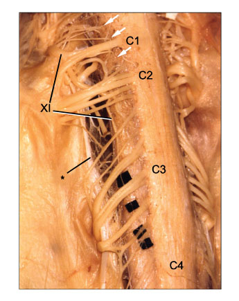
Fig. 3 The most caudal spinal rootlet of the accessory nerve (XI)is at C4. Arrows and asterisk note the spinal rootlets of the accessory nerve at C1 and the denticulate ligament, respectively.
Reference
-
1. Lachman N, Acland R, Rosse C. Anatomical evidence for the absence of a morphologically distinct cranial root of the accessory nerve in man. Clin Anat. 2002. 15:4–10.
Article2. Ryan S, Blyth P, Duggan N, Wild M, Al-Ali S. Is the cranial accessory nerve really a portion of the accessory nerve? Anatomy of the cranial nerves in the jugular foramen. Anat Sci Int. 2007. 82:1–7.
Article3. Clemente CD. Gray's anatomy. 1985. 30th. Philadelphia: Lea & Febiger;1189.4. Romanes GJ. Romanes GJ, editor. The peripheral nervous system. Cunningham's textbook of anatomy. 1981. 12. Oxford: Oxford University Press;764.5. Standring S. Standring S, editor. Ellis H, editor. Healy JC, editor. Johnson D, editor. Williams A, editor. Collins P, editor. Wigley C, editor. Head and neck. Gray's anatomy. 2005. 39th. Edinburgh: Elsevier Churchill Livingstone;558.6. Moore K. Dalley AF. Clinically oriented anatomy. 2006. 5th. Philadelphia: Lippincott Williams & Wilkins;1152.7. Kumaki K. The cervical and the spinal accessory nerves: Morphological studies by means of fibre analysis. Kaibogaku Zasshi. 1970. 45:311–344.8. Holl M. Uber den Nervus accessories Willisii. Arch Anat Phys. 1878. 491–517.9. Weigner K. Beziehungen des nervus accessories zu den proximalen spinalnerven. Anat Hefte 1. Abt. 1901. 17:551–587.10. Uenae F. Über die wurzel der ersten halsnerven des menschen und einiger säugetiere. Acta Sch Med Univ Kioto. 1929. 10:37–49.11. Kim KS, Chung HS, Lee MS. The macroscopic investigation of the spinal cord and accessory nerve in Korean. The Korean Central Journal of Medicine. 1972. 23:309–317.12. Hagenah R, Kosak M, Freckmann N. Anatomic topographical relationship of the intraspinal accessory root to the upper cervical roots and to the vessels of the cranial cervical region. Acta Anat. 1983. 115:158–167.
Article
- Full Text Links
-
- Actions
-
Cited
- CITED
-
- Close
- Share
-
- Similar articles
-
- An Anatomical Study on the Variations of the First Cervical Dorsal Root
- Intradural Variations of Spinal Nerve Rootlets
- A Case of Spinal Accessory Neurilemmoma
- An Immunohistochemical Tracing on the Central Neural Pathways of the Spinal Accessory Nerve using Pseudorabies Virus
- Delayed Spinal Accessory Neuropathy Diagnosed after Local Operative Surgical Procedures in The Posterior Cervical Triangle
