Initial Dead Space and Multiplicity of Bone Flap as Strong Risk Factors for Bone Flap Resorption after Cranioplasty for Traumatic Brain Injury
- Affiliations
-
- 1Department of Neurosurgery, Hallym University Kangnam Sacred Heart Hospital, Hallym University College of Medicine, Seoul, Korea. thlthd@korea.ac.kr
- KMID: 2424321
- DOI: http://doi.org/10.13004/kjnt.2018.14.2.105
Abstract
OBJECTIVE
Bone flap resorption (BFR) is a complication of cranioplasty (CP) that increases the risk of brain damage and can cause cosmetic defects. In this study, the risk factors for BFR were examined to improve the prognosis of patients after CP for traumatic brain injury (TBI).
METHODS
This study was conducted in 80 patients with TBI who underwent decompressive craniectomy and CP with an autologous bone graft between August 2006 and August 2017. BFR was defined as a >0.1 ratio of the difference between the initial bone flap area and the last bone flap area to the craniectomy size and a < 0.5 ratio of the last bone flap thickness to the bone thickness of the contralateral region on computed tomography scans and plain skull radiographs. The patients were divided into the BFR and non-BFR groups, and medical data were compared between the two groups.
RESULTS
Among the 80 patients, 22 (27.5%) were diagnosed as having BFR after CP. The earliest cases of BFR occurred at 57 days after CP, and the latest BFR cases occurred at 3,677 days after CP. Using multivariate logistic regression analyses, the initial dead space size (odds ratio [OR], 1.002; 95% confidence interval [CI], 1.001-1.004; p=0.006) and multiplicity of the bone flap (OR, 3.058; 95% CI, 1.021-9.164; p=0.046) were found to be risk factors for BFR.
CONCLUSION
The risk factors for BFR in this study were the initial dead space size and multiplicity of the bone flap.
Keyword
MeSH Terms
Figure
-
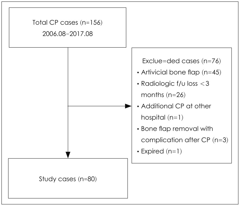
FIGURE 1 Flowchart for the inclusion of patients in this retrospective study. CP: cranioplasty, f/u: follow-up.
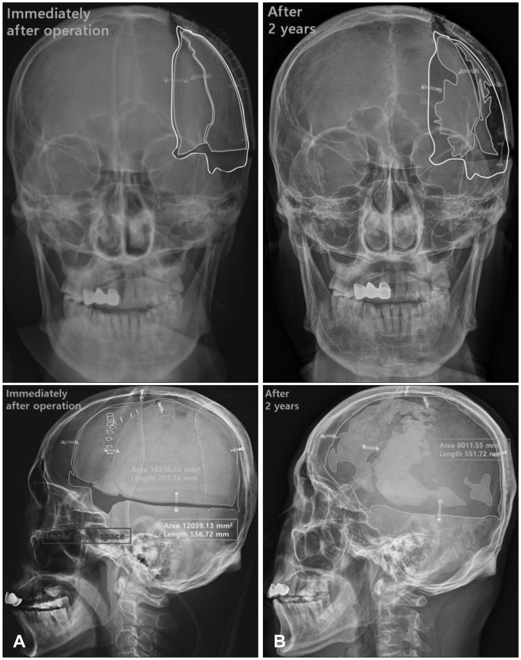
FIGURE 2 Plain skull radiographs in anteroposterior and lateral views. (A) Immediately after the operation; the yellow border indicates the initial bone flap area, the white border indicates the craniectomy area, and the red area indicates the initial dead space. (B) Two years after cranioplasty; the yellow border indicates the last bone flap area, which shows severe bone flap resorption. The areas were measured using free region of interest.
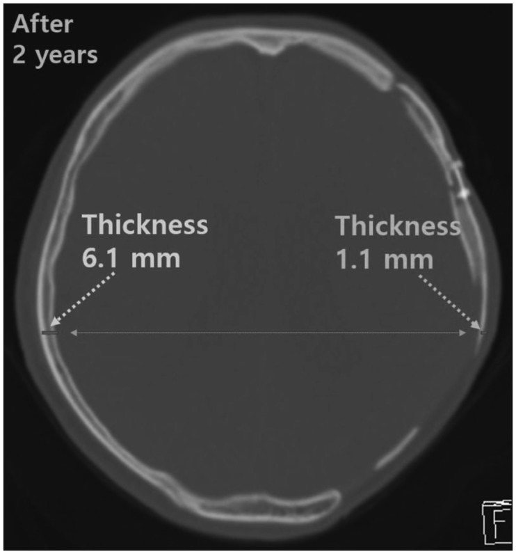
FIGURE 3 Computed tomography image of bone flap resorption at 2 years after cranioplasty, showing partial resorption of the bone flap, in which the remnant bone flap (thickness, 1.1 mm) was <50% as thick as the contralateral region (thickness, 6.1 mm) of the skull.
Reference
-
1. Albrektsson T, Johansson C. Osteoinduction, osteoconduction and osseointegration. Eur Spine J. 2001; 10(Suppl 2):S96–S101. PMID: 11716023.
Article2. Baumeister S, Peek A, Friedman A, Levin LS, Marcus JR. Management of postneurosurgical bone flap loss caused by infection. Plast Reconstr Surg. 2008; 122:195e–208e.
Article3. Bowers CA, Riva-Cambrin J, Hertzler DA 2nd, Walker ML. Risk factors and rates of bone flap resorption in pediatric patients after decompressive craniectomy for traumatic brain injury. J Neurosurg Pediatr. 2013; 11:526–532. PMID: 23473303.
Article4. Brommeland T, Rydning PN, Pripp AH, Helseth E. Cranioplasty complications and risk factors associated with bone flap resorption. Scand J Trauma Resusc Emerg Med. 2015; 23:75. PMID: 26437934.
Article5. Chang V, Hartzfeld P, Langlois M, Mahmood A, Seyfried D. Outcomes of cranial repair after craniectomy. J Neurosurg. 2010; 112:1120–1124. PMID: 19612971.
Article6. Dünisch P, Walter J, Sakr Y, Kalff R, Waschke A, Ewald C. Risk factors of aseptic bone resorption: a study after autologous bone flap reinsertion due to decompressive craniotomy. J Neurosurg. 2013; 118:1141–1147. PMID: 23451904.
Article7. Gooch MR, Gin GE, Kenning TJ, German JW. Complications of cranioplasty following decompressive craniectomy: analysis of 62 cases. Neurosurg Focus. 2009; 26:E9.
Article8. Gosain AK, Gosain SA, Sweeney WM, Song LS, Amarante MT. Regulation of osteogenesis and survival within bone grafts to the calvaria: the effect of the dura versus the pericranium. Plast Reconstr Surg. 2011; 128:85–94. PMID: 21399563.
Article9. Grant GA, Jolley M, Ellenbogen RG, Roberts TS, Gruss JR, Loeser JD. Failure of autologous bone-assisted cranioplasty following decompressive craniectomy in children and adolescents. J Neurosurg. 2004; 100:163–168. PMID: 14758944.
Article10. Honeybul S, Ho KM. Long-term complications of decompressive craniectomy for head injury. J Neurotrauma. 2011; 28:929–935. PMID: 21091342.
Article11. Iwama T, Yamada J, Imai S, Shinoda J, Funakoshi T, Sakai N. The use of frozen autogenous bone flaps in delayed cranioplasty revisited. Neurosurgery. 2003; 52:591–596. PMID: 12590683.
Article12. Jho DH, Neckrysh S, Hardman J, Charbel FT, Amin-Hanjani S. Ethylene oxide gas sterilization: a simple technique for storing explanted skull bone. Technical note. J Neurosurg. 2007; 107:440–445. PMID: 17695404.13. Kalfas IH. Principles of bone healing. Neurosurg Focus. 2001; 10:E1.
Article14. Kim JS, Cheong JH, Ryu JI, Kim JM, Kim CH. Bone flap resorption following cranioplasty after decompressive craniectomy: Preliminary report. Korean J Neurotrauma. 2015; 11:1–5. PMID: 27169057.
Article15. Kim SP, Kang DS, Cheong JH, Kim JH, Song KY, Kong MH. Clinical analysis of epidural fluid collection as a complication after cranioplasty. J Korean Neurosurg Soc. 2014; 56:410–418. PMID: 25535519.
Article16. Klinger DR, Madden C, Beshay J, White J, Gambrell K, Rickert K. Autologous and acrylic cranioplasty: a review of 10 years and 258 cases. World Neurosurg. 2014; 82:e525–e530. PMID: 24036124.
Article17. Martin KD, Franz B, Kirsch M, Polanski W, von der Hagen M, Schackert G, et al. Autologous bone flap cranioplasty following decompressive craniectomy is combined with a high complication rate in pediatric traumatic brain injury patients. Acta Neurochir (Wien). 2014; 156:813–824. PMID: 24532225.
Article18. Park SP, Kim JH, Kang HI, Kim DR, Moon BG, Kim JS. Bone flap resorption following cranioplasty with autologous bone: Quantitative measurement of bone flap resorption and predictive factors. J Korean Neurosurg Soc. 2017; 60:749–754. PMID: 29142636.19. Piedra MP, Thompson EM, Selden NR, Ragel BT, Guillaume DJ. Optimal timing of autologous cranioplasty after decompressive craniectomy in children. J Neurosurg Pediatr. 2012; 10:268–272. PMID: 22861195.
Article20. Piitulainen JM, Kauko T, Aitasalo KM, Vuorinen V, Vallittu PK, Posti JP. Outcomes of cranioplasty with synthetic materials and autologous bone grafts. World Neurosurg. 2015; 83:708–714. PMID: 25681593.
Article21. Redfern RM, Pülhorn H. Cranioplasty. Adv Clin Neurosci Rehabil. 2007; 7:32–34.22. Sanan A, Haines SJ. Repairing holes in the head: a history of cranioplasty. Neurosurgery. 1997; 40:588–603. PMID: 9055300.
Article23. Schoekler B, Trummer M. Prediction parameters of bone flap resorption following cranioplasty with autologous bone. Clin Neurol Neurosurg. 2014; 120:64–67. PMID: 24731578.
Article24. Schuss P, Vatter H, Marquardt G, Imöhl L, Ulrich CT, Seifert V, et al. Cranioplasty after decompressive craniectomy: the effect of timing on postoperative complications. J Neurotrauma. 2012; 29:1090–1095. PMID: 22201297.
Article25. Schwarz F, Dunisch P, Walter J, Sakr Y, Kalff R, Ewald C. Cranioplasty after decompressive craniectomy: is there a rationale for an initial artificial bone-substitute implant? A single-center experience after 631 procedures. J Neurosurg. 2016; 124:710–715. PMID: 26406796.
Article26. Shaffrey ME, Persing JA, Shaffrey CI. Craniofacial reconstruction. In : Apuzzo MLJ, editor. Brain surgery: Complication avoidance and management. New York, NY: Churchill Livingstone;1993. p. 1373–1398.27. Shah AM, Jung H, Skirboll S. Materials used in cranioplasty: a history and analysis. Neurosurg Focus. 2014; 36:E19. PMID: 24684331.
Article28. Wachter D, Reineke K, Behm T, Rohde V. Cranioplasty after decompressive hemicraniectomy: underestimated surgery-associated complications? Clin Neurol Neurosurg. 2013; 115:1293–1297. PMID: 23273384.
Article29. Winkler PA, Stummer W, Linke R, Krishnan KG, Tatsch K. Influence of cranioplasty on postural blood flow regulation, cerebrovascular reserve capacity, and cerebral glucose metabolism. J Neurosurg. 2000; 93:53–61.
Article
- Full Text Links
-
- Actions
-
Cited
- CITED
-
- Close
- Share
-
- Similar articles
-
- Bone Flap Resorption Following Cranioplasty after Decompressive Craniectomy: Preliminary Report
- Analysis of Cranioplasty Using Frozen Autologous Bone Following Post-Traumatic Decompressive Craniectomy
- Risk Factor Analysis of Cryopreserved Autologous Bone Flap Resorption in Adult Patients Undergoing Cranioplasty with Volumetry Measurement Using Conventional Statistics and Machine-Learning Technique
- Bone Flap Resorption Following Cranioplasty with Autologous Bone: Quantitative Measurement of Bone Flap Resorption and Predictive Factors
- Symptomatic Epidural Fluid Collection Following Cranioplasty after Decompressive Craniectomy for Traumatic Brain Injury
