Optimal Dose and Timing of Umbilical Stem Cells Treatment in Pulmonary Arterial Hypertensive Rats
- Affiliations
-
- 1Department of Pediatrics, Ewha Womans University School of Medicine, Seoul, Korea. ymhong@ewha.ac.kr
- 2Department of Thoracic and Cardiovascular Surgery, Ewha Womans University School of Medicine, Seoul, Korea.
- 3Biomedical Research Institute, MEDIPOST, Co., Seoul, Korea.
- KMID: 2419115
- DOI: http://doi.org/10.3349/ymj.2017.58.3.570
Abstract
- PURPOSE
Pulmonary arterial hypertension (PAH) is a fatal disease which is characterized by an increase in pulmonary arterial pressure leading to increases in right ventricular afterload. Human umbilical cord blood derived-mesenchymal stem cells (hUCB-MSCs) administered via the jugular vein have been previously shown to improve PAH by reversal treatment. However, the effect of low dosage and transfusion timing of hUCB-MSCs on PAH has not yet been clearly established. Obviously, low dosage treatment can lead to a reduction in costs. This is the first study on early transfusion effect.
MATERIALS AND METHODS
This study was divided into two parts. The first part is an investigation of dose-dependent effect. hUCB-MSCs were administered into 3 groups of rats (UA: 3×10ⶠcells, UB: 1.5×10ⶠcells, UC: 3×10ⵠcells) via the external jugular vein at week 1 after monocrotaline (MCT) injection. The second part is a search for optimal treatment timing in 3×10ⵠcells dose of hUCB-MSCs administered at day 1 for UD group (low dose of hUCB-MSCs at day 1), at day 1 and week 1 for the UE group (dual transfusion of low dose of hUCB-MSCs at day 1 and week 1) and at 1 week for the UF group (reversal treatment of low dose hUCB-MSC at week 1) after MCT injection.
RESULTS
The administration of 3×10ⵠhUCB-MSCs was as effective as the 3×10ⶠdose in decreasing mean right ventricle (RV) pressure and pulmonary pathological changes. Early treatment with hUCB-MSCs improved mean RV pressure, pulmonary pathological changes and heart collagen 3 protein expression levels in PAH.
CONCLUSION
Low-dose early treatment of hUCB-MSCs is as effective as a high dose treatment of hUCB-MSCs in improving PAH although dual or reversal treatment is still more effective.
MeSH Terms
-
Animals
Disease Models, Animal
Familial Primary Pulmonary Hypertension
Humans
Hypertension, Pulmonary/chemically induced/*therapy
Hypertrophy, Right Ventricular/physiopathology
Male
*Mesenchymal Stem Cell Transplantation
Mesenchymal Stromal Cells/*cytology/metabolism
Monocrotaline/toxicity
Pulmonary Artery/pathology
Rats
Rats, Sprague-Dawley
Time Factors
Monocrotaline
Figure
-
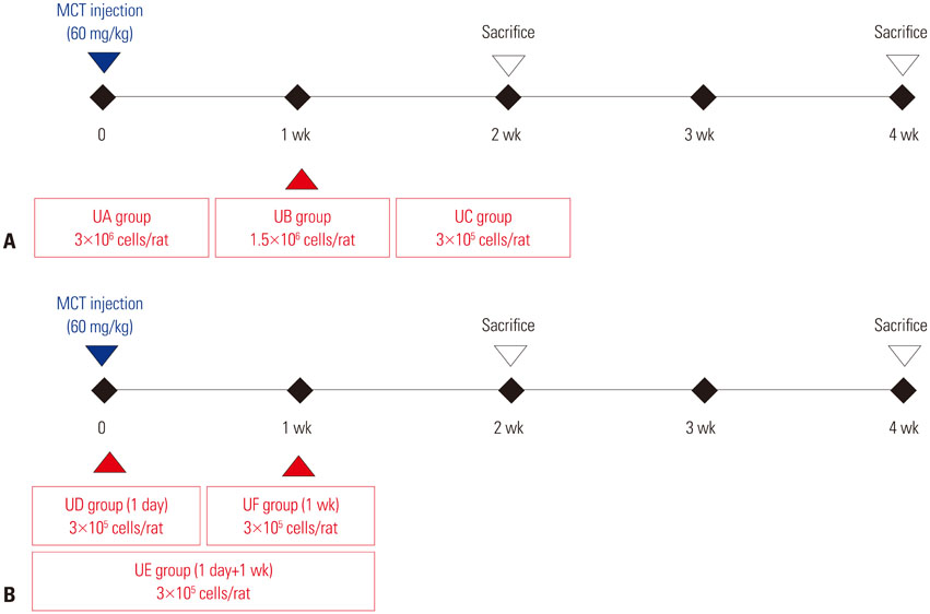
Fig. 1 Study design. (A) Dose-escalation test protocol. The hUCB-MSCs are transfused at 1 week after MCT injection. UA group (n=5) was assigned to receive high-dose hUCB-MSCs (3×10⁶ cells/rat), UB group (n=5) was intermediate-dose hUCB-MSCs (1.5×10⁶ cells/rat) and UC group (n=6) was low-dose hUCB-MSCs (3×10⁵ cells/rat). (B) Treatment time test protocol. Low-dose (3×10⁵ cells/rat) of hUCB-MSCs were transfused at day 1 (UD group, n=4), day 1+week 1 (UE group, n=4) or week 1 (UF group, n=4). MCT, monocrotaline; hUCB-MSCs, human umbilical cord blood derived-mesenchymal stem cells; UD, early treatment of low dose hUCB-MSCs at day 1; UE, dual transfusion of low dose hUCB-MSCs at day 1 and week 1; UF, reversal treatment of low dose hUCB-MSC at week 1.

Fig. 2 Characteristics of hUCB-MSCs. (A and B) The hUCB-MSCs were stained with various combinations of saturating amounts of monoclonal antibodies conjugated with fluorescein isothiocyanate or phycoerythrin: CD14, CD45, HLA-DR, CD73, CD90, and CD105. At least 104 events were analyzed by flow cytometry with the cellquest software. Immunophenotype of hUCB-MSCs. MSC-clone of human cord blood at passage 4 was labeled with antibodies against the indicated antigens and they were analyzed by flow cytometry. FITC-A, isothiocyanate antibody; PE-A, phycoerythrin antibody; hUCB-MSCs, human umbilical cord blood derived-mesenchymal stem cells.
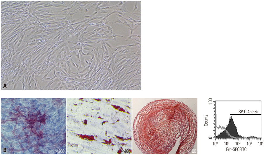
Fig. 3 Differentiation potentiality of hUCB-MSCs on various mesenchymal lineage cells. (A) Inverted phase contrast microscopy images of hUCB-MSCs in the 4 passage. (B) To evaluate the multi-lineage differentiation capacity of the hUCB-MSCs, we cultured them with osteo, adipo, chondrogenic differentiation media. Osteogenic differentiation was observed, demonstrated by morphological changes and the accumulation of alkaline phosphatase. Adipogenic differentiation was observed, shown by the accumulation of oil red-O-staining. Chondrogenic differentiation was also observed, shown by the accumulation of safranin-O-staining sulfated proteoglycans. Finally, differentiation into type II alveolar epithelial cell was observed, shown by SP-C expression. hUCB-MSCs, human umbilical cord blood derived-mesenchymal stem cells.
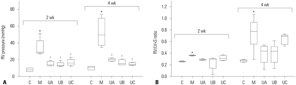
Fig. 4 RV pressure and RV/LV+S ratio in MCT-induced PAH rats according to dose of hUCB-MSCs. (A) RV pressure was significantly decreased in UA, UB and UC groups at weeks 2 and 4. There was no significant difference among the three groups. (B) RV/LV+S ratio slightly decreased in UA, UB and UC groups. *p<0.05, compared with C, †p<0.05, compared with M. MCT, monocrotaline; PAH, pulmonary arterial hypertension; hUCB-MSCs, human umbilical cord blood derived mesenchymal stem cells; RV, right ventricle; LV, left ventricle; S, septum; C, control group; M, monocrotaline group; UA, 3×10⁶ cells/rat; UB, 1.5×10⁶ cells/rat; UC, 3×10⁵ cells/rat.

Fig. 5 Pathologic findings in MCT-induced PAH rats according to dose of hUCB-MSCs at week 4. (A) Low-dose of hUCB-MSCs also effectively decreased pulmonary arteriole medial wall thickening. (B) The number of intra-acinar arteries was decreased in UA and UB groups. (C) Low-dose hUCB-MSCs also attenuated pulmonary pathological changes in MCT-induced PAH rat (Victoria blue staining, ×400). *p<0.05, compared with C, †p<0.05, compared with M. No, number; C, control group; M, monocrotaline group; UA, 3×10⁶ cells/rat; UB, 1.5×10⁶ cells/rat; UC, 3×10⁵ cells/rat; MCT, monocrotaline; PAH, pulmonary arterial hypertension; hUCB-MSCs, human umbilical cord blood derived mesenchymal stem cells.
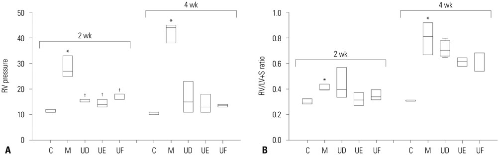
Fig. 6 RV pressure and RV/LV+S ratio according to treatment timing by low-dose treatment of hUCB-MSCs in MCT-induced PAH rats. (A) RV pressure was decreased in UD, UE, and UF group. (B) No effect on RV hypertrophy by low-dose treatment of hUCB-MSCs in MCT-induced PAH rats. *p<0.05, compared with C, †p<0.05, compared with M. MCT, monocrotaline; PAH, pulmonary arterial hypertension; hUCB-MSCs, human umbilical cord blood derived mesenchymal stem cells; RV, right ventricle; LV+S, left ventricle+septum; C, control group; M, monocrotaline group; UD, early treatment group of low dose hUCB-MSCs at day 1; UE, dual transfusion group of low dose hUCB-MSCs at day 1 and week 1; UF, reversal treatment group of low dose hUCB-MSC at week 1; hUCB-MSCs, human umbilical cord blood derived-mesenchymal stem cells.
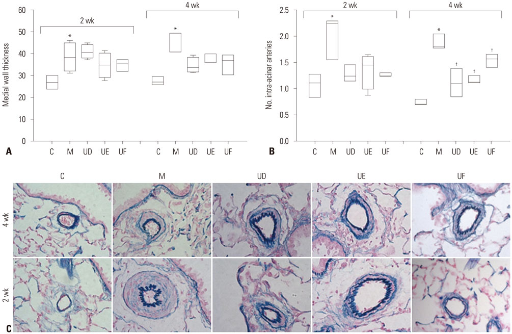
Fig. 7 Pulmonary pathologic finding according to treatment timing by low-dose treatment of hUCB-MSCs in MCT-induced PAH rats. (A) Medial wall thickening of pulmonary arterioles was decreased in UD, UE, and UF groups at week 4, but not significant. (B) The number of mucularized intra-acinar arteries was significantly decreased in UD, UE, and UF groups at weeks 4. (C) Low-dose hUCB-MSCs attenuated pulmonary pathological changes in MCT-induced PAH rat (Victoria blue staining, ×400). *p<0.05, compared with C, †p<0.05, compared with M. MCT, monocrotaline; PAH, pulmonary arterial hypertension; hUCB-MSCs, human umbilical cord blood derived mesenchymal stem cells; C, control group; M, monocrotaline group; UD, early treatment group of low dose hUCB-MSCs at day 1; UE, dual transfusion group of low dose hUCB-MSCs at day 1 and week 1; UF, reversal treatment group of low dose hUCB-MSC at week 1.
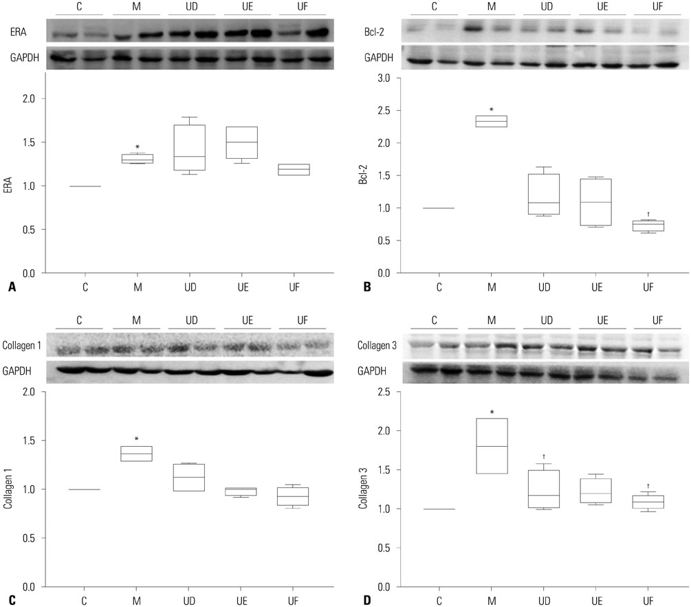
Fig. 8 Expressions of ERA, Bcl-2, collagen 1, and collagen 3 according to treatment timing by low-dose treatment of hUCB-MSCs in MCT-induced PAH rats. (A) Protein expression of ERA was significantly increased in the lung tissues of M group compared with C group at week 4. (B) Protein expression of Bcl-2 was significantly decreased in the lung tissues of UF groups compared with M group at week 4. (C) Collagen 1 in the heart tissues was significantly increased in M group compared with C group at week 4. (D) Collagen 3 in the heart tissues was significantly decreased UD and UF group compared with M group at week 4. *p<0.05, compared with C, †p<0.05, compared with M. MCT, monocrotaline; PAH, pulmonary arterial hypertension; hUCB-MSCs, human umbilical cord blood derived mesenchymal stem cells; C, control group; M, monocrotaline group; UD, early treatment group of low dose hUCB-MSCs at day 1; UE, dual transfusion group of low dose hUCB-MSCs at day 1 and week 1; UF, reversal treatment group of low dose hUCB-MSC at week 1; Dual, twice-transfusion of hUCB-MSCs group; ERA, endothelin receptor A; Bcl-2, B-cell lymphoma 2; GAPDH, glyceraldehyde 3-phosphate dehydrogenase.
Cited by 1 articles
-
Stem Cell and Exosome Therapy in Pulmonary Hypertension
Seyeon Oh, Ji-Hye Jung, Kyung-Jin Ahn, Albert Youngwoo Jang, Kyunghee Byun, Phillip C. Yang, Wook-Jin Chung
Korean Circ J. 2022;52(2):110-122. doi: 10.4070/kcj.2021.0191.
Reference
-
1. Simonneau G, Gatzoulis MA, Adatia I, Celermajer D, Denton C, Ghofrani A, et al. Updated clinical classification of pulmonary hypertension. J Am Coll Cardiol. 2013; 62:25 Suppl. D34–D41.
Article2. Rabinovitch M. Molecular pathogenesis of pulmonary arterial hypertension. J Clin Invest. 2012; 122:4306–4313.
Article3. Huertas A, Perros F, Tu L, Cohen-Kaminsky S, Montani D, Dorfmüller P, et al. Immune dysregulation and endothelial dysfunction in pulmonary arterial hypertension: a complex interplay. Circulation. 2014; 129:1332–1340.
Article4. Price LC, Wort SJ, Perros F, Dorfmüller P, Huertas A, Montani D, et al. Inflammation in pulmonary arterial hypertension. Chest. 2012; 141:210–221.
Article5. Humbert M, Sitbon O, Simonneau G. Treatment of pulmonary arterial hypertension. N Engl J Med. 2004; 351:1425–1436.
Article6. Lee H, Lee JC, Kwon JH, Kim KC, Cho MS, Yang YS, et al. The effect of umbilical cord blood derived mesenchymal stem cells in monocrotaline-induced pulmonary artery hypertension rats. J Korean Med Sci. 2015; 30:576–585.
Article7. Kim KC, Lee JC, Lee H, Cho MS, Choi SJ, Hong YM. Changes in caspase-3, B cell leukemia/lymphoma-2, interleukin-6, tumor necrosis factor-α and vascular endothelial growth factor gene expression after human umbilical cord blood derived mesenchymal stem cells transfusion in pulmonary hypertension rat models. Korean Circ J. 2016; 46:79–92.
Article8. Li J, Li D, Liu X, Tang S, Wei F. Human umbilical cord mesenchymal stem cells reduce systemic inflammation and attenuate LPSinduced acute lung injury in rats. J Inflamm (Lond). 2012; 9:33.
Article9. Hansmann G, Fernandez-Gonzalez A, Aslam M, Vitali SH, Martin T, Mitsialis SA, et al. Mesenchymal stem cell-mediated reversal of bronchopulmonary dysplasia and associated pulmonary hypertension. Pulm Circ. 2012; 2:170–181.
Article10. Chang YS, Ahn SY, Yoo HS, Sung SI, Choi SJ, Oh W, et al. Mesenchymal stem cells for bronchopulmonary dysplasia: phase 1 dose-escalation clinical trial. J Pediatr. 2014; 164:966–972.
Article11. Zhang WG, He L, Shi XM, Wu SS, Zhang B, Mei L, et al. Regulation of transplanted mesenchymal stem cells by the lung progenitor niche in rats with chronic obstructive pulmonary disease. Respir Res. 2014; 15:33.
Article12. Jin Z, Pan X, Zhou K, Bi H, Wang L, Yu L, et al. Biological effects and mechanisms of action of mesenchymal stem cell therapy in chronic obstructive pulmonary disease. J Int Med Res. 2015; 43:303–310.
Article13. Barczyk M, Schmidt M, Mattoli S. Stem cell-based therapy in idiopathic pulmonary fibrosis. Stem Cell Rev. 2015; 11:598–620.
Article14. Weiss DJ. Concise review: current status of stem cells and regenerative medicine in lung biology and diseases. Stem Cells. 2014; 32:16–25.
Article15. Souidi N, Stolk M, Seifert M. Ischemia-reperfusion injury: beneficial effects of mesenchymal stromal cells. Curr Opin Organ Transplant. 2013; 18:34–43.16. Xue J, Li X, Lu Y, Gan L, Zhou L, Wang Y, et al. Gene-modified mesenchymal stem cells protect against radiation-induced lung injury. Mol Ther. 2013; 21:456–465.
Article17. Wannemuehler TJ, Manukyan MC, Brewster BD, Rouch J, Poynter JA, Wang Y, et al. Advances in mesenchymal stem cell research in sepsis. J Surg Res. 2012; 173:113–126.
Article18. Baber SR, Deng W, Master RG, Bunnell BA, Taylor BK, Murthy SN, et al. Intratracheal mesenchymal stem cell administration attenuates monocrotaline-induced pulmonary hypertension and endothelial dysfunction. Am J Physiol Heart Circ Physiol. 2007; 292:H1120–H1128.
Article19. Ionescu L, Byrne RN, van Haaften T, Vadivel A, Alphonse RS, Rey-Parra GJ, et al. Stem cell conditioned medium improves acute lung injury in mice: in vivo evidence for stem cell paracrine action. Am J Physiol Lung Cell Mol Physiol. 2012; 303:L967–L977.
Article20. Lee JW, Fang X, Krasnodembskaya A, Howard JP, Matthay MA. Concise review: mesenchymal stem cells for acute lung injury: role of paracrine soluble factors. Stem Cells. 2011; 29:913–919.
Article21. Umar S, de Visser YP, Steendijk P, Schutte CI, Laghmani EH, Wagenaar GT, et al. Allogenic stem cell therapy improves right ventricular function by improving lung pathology in rats with pulmonary hypertension. Am J Physiol Heart Circ Physiol. 2009; 297:H1606–H1616.
Article22. Kern S, Eichler H, Stoeve J, Klüter H, Bieback K. Comparative analysis of mesenchymal stem cells from bone marrow, umbilical cord blood, or adipose tissue. Stem Cells. 2006; 24:1294–1301.
Article23. Musina RA, Bekchanova ES, Belyavskii AV, Grinenko TS, Sukhikh GT. Umbilical cord blood mesenchymal stem cells. Bull Exp Biol Med. 2007; 143:127–131.
Article24. Liu CH, Hwang SM. Cytokine interactions in mesenchymal stem cells from cord blood. Cytokine. 2005; 32:270–279.
Article25. Lee HR, Kim TH, Choi KJ, Choi KC. Effects of octylphenol on the expression of cell cycle-related genes and the growth of mesenchymal stem cells derived from human umbilical cord blood. Int J Mol Med. 2014; 33:221–226.
Article26. Sitbon O, Humbert M, Nunes H, Parent F, Garcia G, Hervé P, et al. Long-term intravenous epoprostenol infusion in primary pulmonary hypertension: prognostic factors and survival. J Am Coll Cardiol. 2002; 40:780–788.
Article27. Ortiz LA, Gambelli F, McBride C, Gaupp D, Baddoo M, Kaminski N, et al. Mesenchymal stem cell engraftment in lung is enhanced in response to bleomycin exposure and ameliorates its fibrotic effects. Proc Natl Acad Sci U S A. 2003; 100:8407–8411.
Article28. Le Blanc K, Rasmusson I, Sundberg B, Götherström C, Hassan M, Uzunel M, et al. Treatment of severe acute graft-versus-host disease with third party haploidentical mesenchymal stem cells. Lancet. 2004; 363:1439–1441.
Article29. Gitelman SE, Haller MJ, Schatz D. Autologous nonmyeloablative hematopoietic stem cell transplantation in newly diagnosed type 1 diabetes mellitus. JAMA. 2009; 302:624–625.
Article30. Couri CE, Oliveira MC, Stracieri AB, Moraes DA, Pieroni F, Barros GM, et al. C-peptide levels and insulin independence following autologous nonmyeloablative hematopoietic stem cell transplantation in newly diagnosed type 1 diabetes mellitus. JAMA. 2009; 301:1573–1579.
Article31. Mei SH, McCarter SD, Deng Y, Parker CH, Liles WC, Stewart DJ. Prevention of LPS-induced acute lung injury in mice by mesenchymal stem cells overexpressing angiopoietin 1. PLoS Med. 2007; 4:e269.
Article32. Polchert D, Sobinsky J, Douglas G, Kidd M, Moadsiri A, Reina E, et al. IFN-gamma activation of mesenchymal stem cells for treatment and prevention of graft versus host disease. Eur J Immunol. 2008; 38:1745–1755.
Article33. Zhao YD, Courtman DW, Deng Y, Kugathasan L, Zhang Q, Stewart DJ. Rescue of monocrotaline-induced pulmonary arterial hypertension using bone marrow-derived endothelial-like progenitor cells: efficacy of combined cell and eNOS gene therapy in established disease. Circ Res. 2005; 96:442–450.
Article34. Pierro M, Ionescu L, Montemurro T, Vadivel A, Weissmann G, Oudit G, et al. Short-term, long-term and paracrine effect of human umbilical cord-derived stem cells in lung injury prevention and repair in experimental bronchopulmonary dysplasia. Thorax. 2013; 68:475–484.
Article35. van Haaften T, Byrne R, Bonnet S, Rochefort GY, Akabutu J, Bouchentouf M, et al. Airway delivery of mesenchymal stem cells prevents arrested alveolar growth in neonatal lung injury in rats. Am J Respir Crit Care Med. 2009; 180:1131–1142.
Article36. Gnecchi M, Zhang Z, Ni A, Dzau VJ. Paracrine mechanisms in adult stem cell signaling and therapy. Circ Res. 2008; 103:1204–1219.
Article
- Full Text Links
-
- Actions
-
Cited
- CITED
-
- Close
- Share
-
- Similar articles
-
- Stem Cell Transplantation in Umbilical Cord Blood(I) Expansion Effects of Stem Cells in Umbilical Cord Blood with Various Hematopoietic Growth Factors
- Microarray analysis after umbilical cord blood derived mesenchymal stem cells injection in monocrotaline-induced pulmonary artery hypertension rats
- Differentiation of Osteoblast Progenitor Cells from Human Umbilical Cord Blood
- Stem Cell Therapy for Bronchopulmonary Dysplasia: Bench to Bedside Translation
- Establishment of High Throughput Screening System Using Human Umbilical Cord-derived Mesenchymal Stem Cells
