J Adv Prosthodont.
2009 Nov;1(3):118-123.
Evaluation of proximal contact strength by postural changes
- Affiliations
-
- 1Department of Prosthodontics, School of Dentistry, Chosun University, Gwangju, Korea. shoh@chosun.ac.kr
Abstract
- STATEMENT OF PROBLEM: Proper proximal contact is important for maintaining and stabilizing the dental arch. However, the proximal contact strength (PCS) is not a constant value and can be affected by a variety of factors. PURPOSE: This study examined the influences of postural changes on the posterior PCS. MATERIAL AND METHODS: Twelve adults with a normal occlusion and had not undergone prosthetic treatment or proximal restoration were participated in this study. A metal strip was inserted into the proximal surface and removed at a constant velocity. The contact strength was measured in every contact point between canine to second molar in both arches. The PCSs were obtained initially in the upright position, secondly in the supine position and finally in the upright position again. All measurements were repeated after a 2 hour period. Statistical analysis was carried out using the Friedman test (P < .05). RESULTS: Generally, a decrease in PCS occurred when the posture was changed from the initial upright to supine position, while it increased when the posture was changed from the supine to upright position. A significant change was observed in all areas except for between the canine-first premolar in the maxilla and between the first molarsecond molar in the mandible areas. CONCLUSION: The posterior PCS, which dentists generally believe to be a static feature of occlusion, is affected significantly by posture.
Figure
-
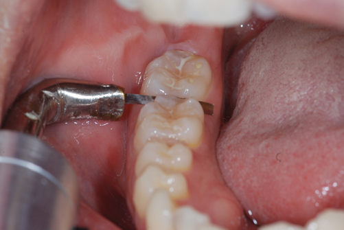
Fig. 1 Measurement of the proximal contact strength between the first molar and second molar in the right side of the mandible.

Fig. 2 Diagrammatic presentation of the changes in proximal contact strength (N) in each region of the maxilla according to the postural change.
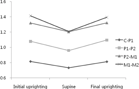
Fig. 3 Diagrammatic presentation of the changes in proximal contact strength (N) in each region of the mandible according to the postural change.
Reference
-
1. The academy of prosthodontics. The glossary of prosthodontic terms. 8th ed. J Prosthet Dent. 2005. 94:37. 57.2. Andrews LF. The six keys to normal occlusion. Am J Orthod. 1972. 62:296–309.3. Wheeler RC. An atlas of tooth form. 1969. 4th ed. Philadelphia, W.B.: Saunders Co;12.4. Sluder TB. Studevant CM, editor. Clinical dental anatomy, histology, physiology and occlusion. The art and science of operative dentistry. 1985. 2nd ed. New York: McGraw-Hill;21.5. Hirschfeld I. Food impaction. J Am Dent Assoc. 1930. 17:1504–1528.6. Hancock EB, Mayo CV, Schwab RR, Wirthlin MR. Influence of interdental contacts on periodontal status. J Periodontol. 1980. 51:445–449.7. Nielsen IM, Glavind L, Karring T. Interproximal periodontal intrabony defects. Prevalence, localization and etiological factors. J Clin Periodontol. 1980. 7:187–198.8. Jernberg GR, Bakdash MB, Keenan KM. Relationship between proximal tooth open contacts and periodontal disease. J Periodontol. 1983. 54:529–533.9. Vardimon AD, Matsaev E, Lieberman M, Brosh T. Tightness of dental contact points in spaced and non-spaced permanent dentitions. Eur J Orthod. 2001. 23:305–314.10. Prakki A, Cilli R, Saad JO, Rodrigues JR. Clinical evaluation of proximal contacts of Class II esthetic direct restorations. Quintessence Int. 2004. 35:785–789.11. Kasahara K, Miura H, Kuriyama M, Kato H, Hasegawa S. Observations of interproximal contact relations during clenching. Int J Prosthodont. 2000. 13:289–294.12. Campagni WV. The final touch in the delivery of a fixed prosthesis. CDA J. 1984. 12:21–29.13. Sturdevant JR, Sturdevant CM. Sturdevant C, Barton R, Sockwell C, Strickland W, editors. Gold inlay and gold onlay restoration for Class II cavity preparations. The Art and Science of Operative Dentistry. 1985. St. Louis, MO, USA: C.V. Mosby Co;490.14. Osborn JW. An investigation into the interdental forces occurring between the teeth of the same arch during clenching the jaws. Arch Oral Biol. 1961. 5:202–211.15. Dörfer CE, von Bethlenfalvy ER, Staehle HJ, Pioch T. Factors influencing proximal dental contact strengths. Eur J Oral Sci. 2000. 108:368–377.16. Oh SH, Nakano M, Bando E, Keisuke N, Shigemoto S, Jeong JH, Kang DW. Relationship between occlusal tooth contact patterns and tightness of proximal tooth contact. J Oral Rehabil. 2006. 33:749–753.17. Southard TE, Behrents RG, Tolley EA. The anterior component of occlusal force. Part 1. Measurement and distribution. Am J Orthod Dentofacial Orthop. 1989. 96:493–500.18. Choi WJ, Kim KH, Kim JA, Kang DW, Oh SH. Evaluation and development of digital device for measuring proximal tooth contact tightness. J Korean Acad Prosthodont. 2007. 45:687–695.19. Southard TE, Southard KA, Tolley EA. Variation of approximal tooth contact tightness with postural change. J Dent Res. 1990. 69:1776–1779.20. Shames IH. Engineering Mechanics. 1966. Vol. 1. Englewood Cliffs, NJ: Prentice-Hall;170–174.21. Fuhrmann R, Grave C, Diedrich P. In vitro evaluation of a measurement method to analyze the interdental, mesially directed force. J Orofac Orthop. 1998. 59:362–370.22. Kim KH. Evaluation of proximal tooth contact tightness in permanent dentitions. 2007. Korea: Chosun University School of Dentistry;M.S. Thesis.23. Southard TE, Southard KA, Tolley EA. Periodontal force: a potential cause of relapse. Am J Orthod Dentofacial Orthop. 1992. 101:221–227.24. Miyamoto K, Yamada K, Ishizuka Y, Morimoto N, Tanne K. Masseter muscle activity during the whole day in young adults. Am J Orthod Dentofacial Orthop. 1996. 110:394–398.25. Smith JJ, Kampine JP. Circulatory physiology- the essential. 1984. Baltimore, MD: Waverly Press, Inc.;235–237.26. Rowell LB, Blackmon JR. Hunyor S, Ludbrook J, Shaw J, McGrath M, editors. Reflex and local control of the splanchnic circulation in humans. The peripheral circulation. 1984. New York: Excerpta Medica;114.27. Moxham BJ. Davidovitch Z, editor. The role of the periodontal vasculature in tooth eruption. The biological mechanisms of tooth eruption and root resorption. 1988. Birmingham: EBSCO Media;208.28. Körber KH. Periodontal pulsation. J Periodontol. 1970. 41:382–390.29. Ng GC, Walker TW, Zingg W, Burke PS. Effects of tooth loading on the periodontal vasculature of the mandibular fourth premolar in dogs. Arch Oral Biol. 1981. 26:189–195.30. Boice PA, Niles SM, Dubois LM. Evaluation of proximal contacts with shim stock. J Oral Rehabil. 1987. 14:91–94.
- Full Text Links
-
- Actions
-
Cited
- CITED
-
- Close
- Share
-
- Similar articles
-
- Evaluation of the proximal contact and comparison of methods for measuring in normal dentition
- Effect of Transcranial Direct Current Stimulation on Postural Stability and Lower Extremity Strength in Hemiplegic Stroke Patients
- Effects of Community-based Comprehensive Fall Prevention Program on Muscle Strength, Postural Balance and Fall Efficacy in Elderly People
- The Factors associated with Postural Control after Anterior Cruciate Ligament Reconstruction
- Hip Extensor Strength Influences Dynamic Postural Changes during Gait in Patients with Adult Spinal Deformity: A Cross-Sectional Study Using Three-Dimensional Motion Analysis
