Korean J Ophthalmol.
2017 Jun;31(3):275-276. 10.3341/kjo.2017.0013.
A Case of Retinal Cavernous Hemangioma Diagnosed with Spectral Domain Optical Coherence Tomography
- Affiliations
-
- 1Department of Ophthalmology, Samsung Medical Center, Sungkyunkwan University School of Medicine, Seoul, Korea. swkang@skku.edu
- KMID: 2379885
- DOI: http://doi.org/10.3341/kjo.2017.0013
Abstract
- No abstract available.
Figure
-
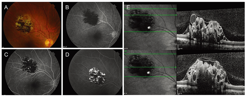
Fig. 1 (A) A color fundus photograph of the right eye of the retinal cavernous hemangioma (patient at the first visit). Whitish-violet color retinal lesion is seen along the superotemporal arcade. (B-D) An early-phase fluorescein angiography image shows that the cystic space unstained with fluorescein, appears hypofluorescent. A late-phase fluorescein angiography image shows that the superior vascular cystic space, which is stained with fluorescein, appears hyperfluorescent. Hypofluorescence in the inferior space is related to erythrocytic sedimentation. (E) Spectral domain optical coherence tomography shows that the ‘grape bunch’ shape cystic hemangioma is located under the internal limiting membrane.
Reference
-
1. Gass JD. Cavernous hemangioma of the retina: a neuro-oculo-cutaneous syndrome. Am J Ophthalmol. 1971; 71:799–814.2. Naftchi S, la Cour M. A case of central visual loss in a child due to macular cavernous haemangioma of the retina. Acta Ophthalmol Scand. 2002; 80:550–552.3. Shields JA, Eagle RC Jr, Ewing MQ, et al. Retinal cavernous hemangioma: fifty-two years of clinical follow-up with clinicopathologic correlation. Retina. 2014; 34:1253–1257.4. Messmer E, Laqua H, Wessing A, et al. Nine cases of cavernous hemangioma of the retina. Am J Ophthalmol. 1983; 95:383–390.5. Pringle E, Chen S, Rubinstein A, et al. Optical coherence tomography in retinal cavernous haemangioma may explain the mechanism of vitreous haemorrhage. Eye (Lond). 2009; 23:1242–1243.
- Full Text Links
-
- Actions
-
Cited
- CITED
-
- Close
- Share
-
- Similar articles
-
- Fundus Autofluorescence, Fluorescein Angiography and Spectral Domain Optical Coherence Tomography Findings of Retinal Astrocytic Hamartomas in Tuberous Sclerosis
- A Case of Ocular Toxoplasmosis Imaged with Spectral Domain Optical Coherence Tomography
- Spectral-Domain Optical Coherence Tomography Findings in Acute Central Retinal Artery Occlusion
- Short-Term Clinical Observation of Acute Retinal Pigment Epitheliitis Using Spectral-Domain Optical Coherence Tomography
- Spectral-domain Optical Coherence Tomography of Combined Hamartoma of the Retina and Retinal Pigment Epithelium in Neurofibromatosis
