Short-Term Clinical Observation of Acute Retinal Pigment Epitheliitis Using Spectral-Domain Optical Coherence Tomography
- Affiliations
-
- 1Department of Ophthalmology, Soonchunhyang University Hospital, Bucheon, Korea. tkpark@schbc.ac.kr
- KMID: 1010034
- DOI: http://doi.org/10.3341/kjo.2011.25.3.222
Abstract
- We investigated the case of a young man with blurred vision in his left eye. His visual acuity was slightly decreased, and ophthalmoscopy disclosed a gray-white lesion in the macula. He had no systemic or ocular history. On the visual field test, the threshold sensitivity was decreased in the corresponding region. Spectral domain optical coherence tomography (OCT) demonstrated a disruption in the photoreceptor inner and outer segment (IS/OS) junction and undulation of the retinal pigment epithelium (RPE) with backscattering. We re-examined the patient after two weeks and after three months without any treatment. Visual acuity and visual field results were gradually normalized, and OCT demonstrated the recovery of continuity in the photoreceptor IS/OS junction, as well as decreased RPE irregularity with minimal backscattering. We used spectral domain OCT instead of time domain OCT (OCT3) so that we could provide better image resolution of the acute retinal pigment epitheliitis (ARPE). Finally, we observed recovery of the functional and anatomical changes in the ARPE patient with a resolution of the condition within three months following the initial examination, using OCT and visual field tests.
MeSH Terms
Figure
-

Fig. 1 Fundus photographs of 18-year-old man. (A) A fundus photograph showed a gray-white lesion in the superior-juxtafoveal region. (B) Three months later, the lesion was diminished in size and intensity.
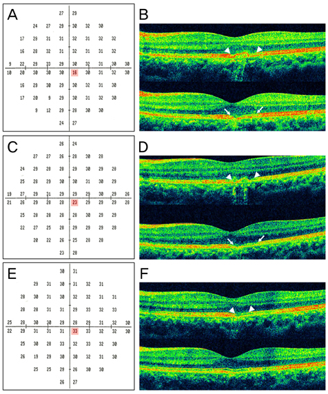
Fig. 2 Eighteen-year-old man: optical coherence tomography (OCT) and visual field. (A) The visual field revealed decreased threshold sensitivity in the macula. (B) OCT demonstrated disruption in the photoreceptor inner and outer segment junction and undulation of the retinal pigment epithelium (RPE) with backscattering. (C) Two weeks after onset, an increased threshold was revealed in the prior point. (D) OCT did not reveal any improvement compared to the initial findings. (E) Three months later, the visual field was normalized. (F) Recovered continuity in the photoreceptor inner and outer segment junction and decreased RPE irregularity with minimal backscattering were demonstrated upon OCT. To determine the serial changes in the lesion, OCT findings were obtained in the identical region of the macula. Acquisition photos were acquired with a five-line raster scan, which consists of five horizontal lines, separated from one other by 0.25 mm. We present only two lines of the raster scan because the other three raster scan lines showed normal findings.
Reference
-
1. Krill AE, Deutman AF. Acute retinal pigment epitheliitus. Am J Ophthalmol. 1972. 74:193–205.2. Chittum ME, Kalina RE. Acute retinal pigment epitheliitis. Ophthalmology. 1987. 94:1114–1119.3. Hsu J, Fineman MS, Kaiser RS. Optical coherence tomography findings in acute retinal pigment epitheliitis. Am J Ophthalmol. 2007. 143:163–165.4. Quillen DA, Davis JB, Gottlieb JL, et al. The white dot syndromes. Am J Ophthalmol. 2004. 137:538–550.5. Li D, Kishi S. Restored photoreceptor outer segment damage in multiple evanescent white dot syndrome. Ophthalmology. 2009. 116:762–770.
- Full Text Links
-
- Actions
-
Cited
- CITED
-
- Close
- Share
-
- Similar articles
-
- Fundus Autofluorescence, Fluorescein Angiography and Spectral Domain Optical Coherence Tomography Findings of Retinal Astrocytic Hamartomas in Tuberous Sclerosis
- Spectral Domain Optical Coherence Tomography Findings of Butterfly Shaped Pigment Dystrophy
- Spectral-Domain Optical Coherence Tomography Findings in Acute Central Retinal Artery Occlusion
- Spectral-domain Optical Coherence Tomography of Combined Hamartoma of the Retina and Retinal Pigment Epithelium in Neurofibromatosis
- Choroidal Thickness at the Outside of Fovea in Diabetic Retinopathy Using Spectral-Domain Optical Coherence Tomography
