Spectral-domain Optical Coherence Tomography of Combined Hamartoma of the Retina and Retinal Pigment Epithelium in Neurofibromatosis
- Affiliations
-
- 1Department of Ophthalmology, Institute of Vision Research, Yonsei University College of Medicine, Seoul, Korea.
- 2Department of Ophthalmology, National Health Insurance Corporation Ilsan Hospital, Goyang, Korea. eunjee95@hanmail.net
- KMID: 1501802
- DOI: http://doi.org/10.3341/kjo.2013.27.1.68
Abstract
- A 5-year-old girl was diagnosed with neurofibromatosis type 2 (NF-2) due to multiple neurofibromas, cafe-au-lait spots, and schwannomas of the brain. During ophthalmologic evaluation, a posterior subcapsular cataract and a gray-green colored subretinal lesion were found in right eye. Fluorescein angiography (FA) revealed a combined hamartoma of the retina and retinal pigment epithelium (CHRRPE). At age 9, she underwent cataract surgery. At this time FA and spectral-domain optical coherence tomography (SD-OCT) were taken. The SD-OCT showed an elevated hyperreflective mass in the retina with prominent attenuation of the inner and outer retina, but minimal attenuation in the photoreceptor layers. The underlying retina appeared to be disorganized and thick (791 microm). This is the first case report of SD-OCT imaging of a CHRRPE associated with NF-2 in a pediatric patient. By using SD-OCT in this patient, we could obtain detailed tumor characteristics, and SD-OCT may be helpful in the diagnosis and management of CHRRPE.
Keyword
MeSH Terms
Figure
-
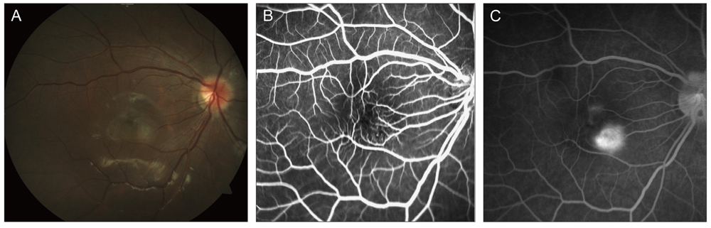
Fig. 1 At age 9, (A) fundus photography of the right eye reveals a moderately pigmented and elevated lesion at the inferotemporal vascular arcade, involving the macula. (B) Fundus fluorescein angiography of the right eye demonstrates some areas of pinpoint hyperfluorescence. (C) Late phase of fluorescein angiography demonstrates mild retinal vascular leakage, and shows no significant progression compared with the previous examination.
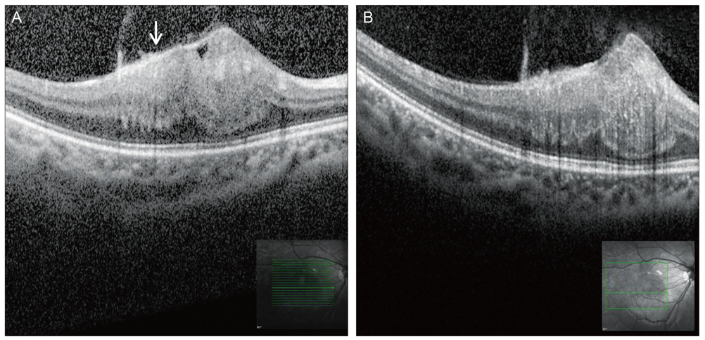
Fig. 2 Spectral-domain optical coherence tomography reveals an elevated hyperreflective mass in the retina with mild attenuation of the retinal pigment epithelium and photoreceptor inner segment/outer segment junction. Prominent thickening and attenuation of the inner retina is also noted (A,B). The arrow in (A) represents the hyperreflective epiretinal membrane.
Reference
-
1. De Laey JJ, Hanssens M. Vascular tumors and malformations of the ocular fundus. 1990. Dordrecht: Kluwer Academic Publishers;101–120.2. Evans DG, Huson SM, Donnai D, et al. A clinical study of type 2 neurofibromatosis. Q J Med. 1992. 84:603–618.3. Evans DG, Huson SM, Donnai D, et al. A genetic study of type 2 neurofibromatosis in the United Kingdom. I. Prevalence, mutation rate, fitness, and confirmation of maternal transmission effect on severity. J Med Genet. 1992. 29:841–846.4. Pearson-Webb MA, Kaiser-Kupfer MI, Eldridge R. Eye findings in bilateral acoustic (central) neurofibromatosis: association with presenile lens opacities and cataracts but absence of Lisch nodules. N Engl J Med. 1986. 315:1553–1554.5. Bouzas EA, Freidlin V, Parry DM, et al. Lens opacities in neurofibromatosis 2: further significant correlations. Br J Ophthalmol. 1993. 77:354–357.6. Landau K, Dossetor FM, Hoyt WF, Muci-Mendoza R. Retinal hamartoma in neurofibromatosis 2. Arch Ophthalmol. 1990. 108:328–329.7. Kaye LD, Rothner AD, Beauchamp GR, et al. Ocular findings associated with neurofibromatosis type II. Ophthalmology. 1992. 99:1424–1429.8. Dossetor FM, Landau K, Hoyt WF. Optic disk glioma in neurofibromatosis type 2. Am J Ophthalmol. 1989. 108:602–603.9. Freedman SF, Elner VM, Donev I, et al. Intraocular neurilemmoma arising from the posterior ciliary nerve in neurofibromatosis: pathologic findings. Ophthalmology. 1988. 95:1559–1564.10. Schachat AP, Shields JA, Fine SL, et al. Combined hamartomas of the retina and retinal pigment epithelium. Ophthalmology. 1984. 91:1609–1615.11. Chan CC, Koch CA, Kaiser-Kupfer MI, et al. Loss of heterozygosity for the NF2 gene in retinal and optic nerve lesions of patients with neurofibromatosis 2. J Pathol. 2002. 198:14–20.12. Hartnett ME, Trese M, Capone A, et al. Pediatric retina: medical and surgical approaches. 2005. Philadelphia: Lippincott Williams and Wilkins;231–233.13. Schachat AP, Shields JA, Fine SL, et al. Combined hamartomas of the retina and retinal pigment epithelium. Ophthalmology. 1984. 91:1609–1615.14. Ryan SJ, Ogden J, Hinton D, Schachat AP. Retina: basic science and inherited retinal disease. 2006. 4th ed. Baltimore: Mosby;673–678.15. Schachat AP, Glaser BM. Retinal hamartoma, acquired retinoschisis, and retinal hole. Am J Ophthalmol. 1985. 99:604–605.16. Kahn D, Goldberg MF, Jednock N. Combined retinal-retina pigment epithelial hamartoma presenting as a vitreous hemorrhage. Retina. 1984. 4:40–43.17. Shields CL, Mashayekhi A, Dai VV, et al. Optical coherence tomographic findings of combined hamartoma of the retina and retinal pigment epithelium in 11 patients. Arch Ophthalmol. 2005. 123:1746–1750.18. Huot CS, Desai KB, Shah VA. Spectral domain optical coherence tomography of combined hamartoma of the retina and retinal pigment epithelium. Ophthalmic Surg Lasers Imaging. 2009. 40:322–324.19. McDonald HR, Abrams GW, Burke JM, Neuwirth J. Clinicopathologic results of vitreous surgery for epiretinal membranes in patients with combined retinal and retinal pigment epithelial hamartomas. Am J Ophthalmol. 1985. 100:806–813.20. Sappenfield DL, Gitter KA. Surgical intervention for combined retinal-retinal pigment epithelial hamartoma. Retina. 1990. 10:119–124.21. Mason JO 3rd. Visual improvement after pars plana vitrectomy and membrane peeling for vitreoretinal traction associated with combined hamartoma of the retina and retinal pigment epithelium. Retina. 2002. 22:824–825.22. Stallman JB. Visual improvement after pars plana vitrectomy and membrane peeling for vitreoretinal traction associated with combined hamartoma of the retina and retinal pigment epithelium. Retina. 2002. 22:101–104.23. Konstantinidis L, Chamot L, Zografos L, Wolfensberger TJ. Pars Plana vitrectomy and epiretinal membrane peeling for vitreoretinal traction associated with combined hamartoma of the retina and retinal pigment epithelium (CHRRPE). Klin Monbl Augenheilkd. 2007. 224:356–359.24. Cohn AD, Quiram PA, Drenser KA, et al. Surgical outcomes of epiretinal membranes associated with combined hamartoma of the retina and retinal pigment epithelium. Retina. 2009. 29:825–830.25. Zhang X, Dong F, Dai R, Yu W. Surgical management of epiretinal membrane in combined hamartomas of the retina and retinal pigment epithelium. Retina. 2010. 30:305–309.
- Full Text Links
-
- Actions
-
Cited
- CITED
-
- Close
- Share
-
- Similar articles
-
- Fundus Autofluorescence, Fluorescein Angiography and Spectral Domain Optical Coherence Tomography Findings of Retinal Astrocytic Hamartomas in Tuberous Sclerosis
- Short-Term Clinical Observation of Acute Retinal Pigment Epitheliitis Using Spectral-Domain Optical Coherence Tomography
- Spectral Domain Optical Coherence Tomography Findings of Butterfly Shaped Pigment Dystrophy
- Choroidal Thickness at the Outside of Fovea in Diabetic Retinopathy Using Spectral-Domain Optical Coherence Tomography
- A Case of Congenital Simple Hamartoma of the Retinal Pigment Epithelium and Coats' Disease in the Same Eye
