The Effect of Distal Hooks in Thoracolumbar Fusion Using a Pedicle Screw in Elderly Patients
- Affiliations
-
- 1Department of Orthopedic Surgery, Inje University Haeundae Paik Hospital, Busan, Korea. sskim@paik.ac.kr
- 2Department of Orthopedic Surgery, Inje University Ilsan Paik Hospital, Goyang, Korea.
- 3Department of Orthopedic Surgery, Inje University Sanggye Paik Hospital, Seoul, Korea.
- KMID: 2369575
- DOI: http://doi.org/10.4055/jkoa.2017.52.1.83
Abstract
- PURPOSE
To investigate the clinical outcomes of distal hook augmentation using a pedicle screw in thoracolumbar fusion in elderly patients.
MATERIALS AND METHODS
This retrospective multicenter study recruited 20 patients aged 65 years or older, who underwent anterior support and long level posterior fusion in the thoracolumbar junction with a follow-up of one year. To assess the effect of distal hook augmentation, the patients were divided into two groups; the pedicle screw with hook group (PH group, n=10) and the pedicle screw alone group (PA group, n=10).
RESULTS
The average age was 72.4 years (65-83 years). The average fusion segment was 4.6 segments (3-6 segments). There were no significant differences in age, sex, causative diseases, bone mineral density of lumbar and proximal femur, number of patients with osteoporosis, and number of fused segments between the two groups (p≥0.05). At 1 year follow-up after surgery, parameters related with distal screw pullout were significantly worse in the PA group. No patients in the PH group had distal screw pullout. However, six patients (60%, 6/10) in the PA group had distal screw pullout. There were no significant differences in the progression of distal junctional kyphosis between the two groups.
CONCLUSION
Distal hook augmentation is an effective procedure in protecting distal pedicle screws against the pullout when long level thoracolumbar fusion was performed in elderly patients aged 65 years or older.
Keyword
MeSH Terms
Figure
-
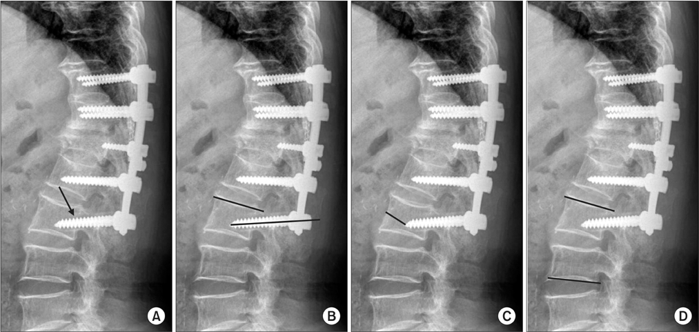
Figure 1 Radiographic parameters. (A) The halo around the distal screw was checked by a radiolucent width from the screw thread end (arrow). (B) The insertion angle of the distal screw was measured by a Cobb angle between the upper endplate of the lowest instrumented vertebrae and its screw. (C) The insertion distance of the distal screw was measured by the distance from the midpoint of anterior cortex of the lowest instrumented vertebrae to its screw tip. (D) Distal junctional lordosis was measured by a Cobb angle between the upper end plate of the lowest instrumented vertebrae and the low end plate of the distal adjacent vertebrae.
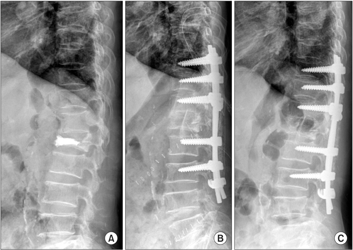
Figure 2 A 77-year-old female patient with distal hook augmentation. She had had back pain and paresthesia on the lower extremities for four months and underwent vertebroplasty for L1 compression fracture at a local clinic two months ago. However, her symptoms were not relieved. She was finally diagnosed with tuberculosis spondylodiscitis. (A) Initial lateral radiography showed bone cement at L1. (B) She was treated by an anterior support using an autoiliac strut bone graft and posterior fusion from T10 to L3 using a pedicle screw with distal laminar hooks. (C) At the 1-year follow-up, there was no pullout of the distal screw.
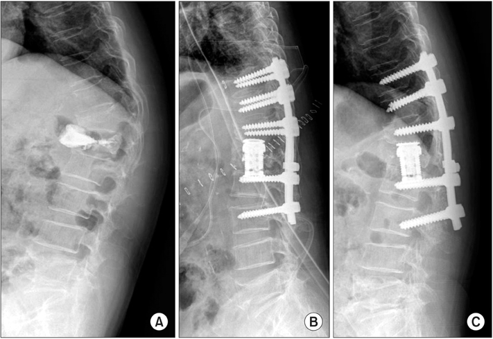
Figure 3 A 70-year-old female patient treated without distal hook augmentation. She had severe back pain and paresthesia on both lower extremities after L1 kyphoplasty at a local clinic six months ago. (A) The cement mass was separated and migrated anteriorly with increasing kyphotic deformity. (B) She was treated by an anterior support using expandable cage and posterior fusion from T10 to L3 using a pedicle screw without distal hook. (C) At the 1-year follow-up, the distal screw was pulled out.
Reference
-
1. Cordista A, Conrad B, Horodyski M, Walters S, Rechtine G. Biomechanical evaluation of pedicle screws versus pedicle and laminar hooks in the thoracic spine. Spine J. 2006; 6:444–449.
Article2. Hasegawa K, Takahashi HE, Uchiyama S, et al. An experimental study of a combination method using a pedicle screw and laminar hook for the osteoporotic spine. Spine (Phila Pa 1976). 1997; 22:958–962. discussion 963.
Article3. Paxinos O, Tsitsopoulos PP, Zindrick MR, et al. Evaluation of pullout strength and failure mechanism of posterior instrumentation in normal and osteopenic thoracic vertebrae. J Neurosurg Spine. 2010; 13:469–476.
Article4. Tai CL, Chen LH, Lee DM, Liu MY, Lai PL. Biomechanical comparison of different combinations of hook and screw in one spine motion unit--an experiment in porcine model. BMC Musculoskelet Disord. 2014; 15:197.
Article5. Tan JS, Kwon BK, Dvorak MF, Fisher CG, Oxland TR. Pedicle screw motion in the osteoporotic spine after augmentation with laminar hooks, sublaminar wires, or calcium phosphate cement: a comparative analysis. Spine (Phila Pa 1976). 2004; 29:1723–1730.
Article6. Glassman SD, Alegre GM. Adult spinal deformity in the osteoporotic spine: options and pitfalls. Instr Course Lect. 2003; 52:579–588.7. Fujita T, Kostuik JP, Huckell CB, Sieber AN. Complications of spinal fusion in adult patients more than 60 years of age. Orthop Clin North Am. 1998; 29:669–678.
Article8. Burval DJ, McLain RF, Milks R, Inceoglu S. Primary pedicle screw augmentation in osteoporotic lumbar vertebrae: biomechanical analysis of pedicle fixation strength. Spine (Phila Pa 1976). 2007; 32:1077–1083.9. Hilibrand AS, Moore DC, Graziano GP. The role of pediculolaminar fixation in compromised pedicle bone. Spine (Phila Pa 1976). 1996; 21:445–451.
Article10. Leduc S, Mac-Thiong JM, Maurais G, Jodoin A. Posterior pedicle screw fixation with supplemental laminar hook fixation for the treatment of thoracolumbar burst fractures. Can J Surg. 2008; 51:35–40.11. Abshire BB, McLain RF, Valdevit A, Kambic HE. Characteristics of pullout failure in conical and cylindrical pedicle screws after full insertion and back-out. Spine J. 2001; 1:408–414.
Article12. Cho W, Cho SK, Wu C. The biomechanics of pedicle screwbased instrumentation. J Bone Joint Surg Br. 2010; 92:1061–1065.
Article13. Suzuki T, Abe E, Okuyama K, Sato K. Improving the pullout strength of pedicle screws by screw coupling. J Spinal Disord. 2001; 14:399–403.
Article14. Karakaşlı A, Sekik E, Karaarslan A, Kızmazoğlu C, Havıtçoğlu H. Are pedicular screws and lateral hook screws more resistant against pullout than conventional spinal hooks and screws in terminal vertebral segment fixation? Eklem Hastalik Cerrahisi. 2016; 27:22–28.
Article15. Kaymaz B, Demirkiran G, Ayvaz M, Akel I, Acaroğlu E, Alanay A. Treatment of thoracolumbar burst fractures using combined pedicle screw-laminar hook fixation. Acta Orthop Traumatol Turc. 2014; 48:152–156.
Article16. Sun E, Alkalay R, Vader D, Snyder BD. Preventing distal pullout of posterior spine instrumentation in thoracic hyperkyphosis: a biomechanical analysis. J Spinal Disord Tech. 2009; 22:270–277.17. Sandén B, Olerud C, Petrén-Mallmin M, Johansson C, Larsson S. The significance of radiolucent zones surrounding pedicle screws. Definition of screw loosening in spinal instrumentation. J Bone Joint Surg Br. 2004; 86:457–461.18. Gruskay JA, Webb ML, Grauer JN. Methods of evaluating lumbar and cervical fusion. Spine J. 2014; 14:531–539.
Article19. Sacramento-Domínguez C, Vayas-Díez R, Coll-Mesa L, et al. Reproducibility measuring the angle of proximal junctional kyphosis using the first or the second vertebra above the upper instrumented vertebrae in patients surgically treated for scoliosis. Spine (Phila Pa 1976). 2009; 34:2787–2791.
Article20. Hirano T, Hasegawa K, Takahashi HE, et al. Structural characteristics of the pedicle and its role in screw stability. Spine (Phila Pa 1976). 1997; 22:2504–2509. discussion 2510.
Article21. You JW, Lim TH. Biomechanical evaluation of supplemental hook or screw fixation in short segment spinal instrumentation. J Korean Soc Spine Surg. 1998; 1:1–8.22. Hitchon PW, Brenton MD, Black AG, et al. In vitro biomechanical comparison of pedicle screws, sublaminar hooks, and sublaminar cables. J Neurosurg. 2003; 99:1 Suppl. 104–109.
Article23. Park P, Garton HJ, Gala VC, Hoff JT, McGillicuddy JE. Adjacent segment disease after lumbar or lumbosacral fusion: review of the literature. Spine (Phila Pa 1976). 2004; 29:1938–1944.
Article24. Shea TM, Laun J, Gonzalez-Blohm SA, et al. Designs and techniques that improve the pullout strength of pedicle screws in osteoporotic vertebrae: current status. Biomed Res Int. 2014; 2014:748393.
Article25. Ferguson RL, Tencer AF, Woodard P, Allen BL Jr. Biomechanical comparisons of spinal fracture models and the stabilizing effects of posterior instrumentations. Spine (Phila Pa 1976). 1988; 13:453–460.
Article26. Ashman RB, Bechtold JE, Edwards WT, Johnston CE 2nd, McAfee PC, Tencer AF. In vitro spinal arthrodesis implant mechanical testing protocols. J Spinal Disord. 1989; 2:274–281.
Article27. Ashman RB, Birch JG, Bone LB, et al. Mechanical testing of spinal instrumentation. Clin Orthop Relat Res. 1988; 227:113–125.
Article
- Full Text Links
-
- Actions
-
Cited
- CITED
-
- Close
- Share
-
- Similar articles
-
- Surgical Management of Thoracolumbar Spine Fracture with Pedicle Screws and Inferior Laminar Hooks
- Surgical Treatment of Congential Scoliosis-Validity of Pedicle Screws
- Comparison of Posterior Fixation Alone and Supplementation with Posterolateral Fusion in Thoracolumbar Burst Fractures
- Segmental Pedicle Screw Fixation in Thoracolumbar or Lumbar Idiopathic Scoliosis
- Change of Canal Compromise After Ligamentotaxis in Thoracolumbar Burst Fracture
