J Pathol Transl Med.
2017 Jan;51(1):7-8. 10.4132/jptm.2016.10.26.
Perivascular Epithelioid Cell Tumors (PEComas) of the Orbit
- Affiliations
-
- 1Department of Surgical, Microsurgical and Medical Sciences, University of Sassari, Sassari, Italy. panospaliogiannis@gmail.com
- 2Institute of Biomolecular Chemistry, Cancer Genetics Unit, C.N.R., Sassari, Italy.
- KMID: 2367674
- DOI: http://doi.org/10.4132/jptm.2016.10.26
Abstract
- No abstract available.
Figure
-
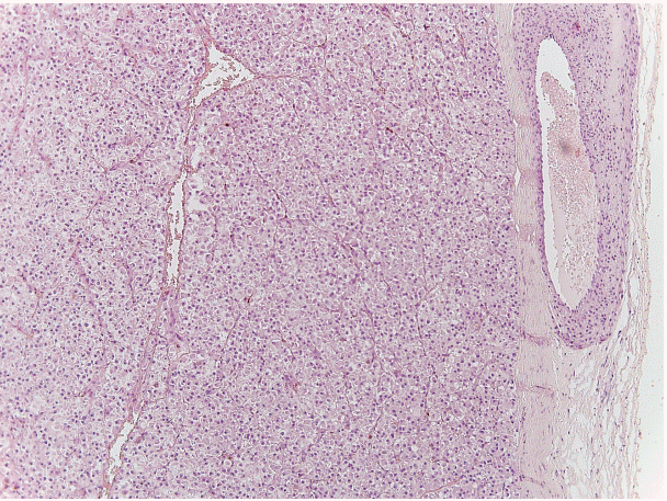
Fig. 1. A section of the lesion stained with hematoxylin and eosin, evidencing its microscopic features and perivascular origin.
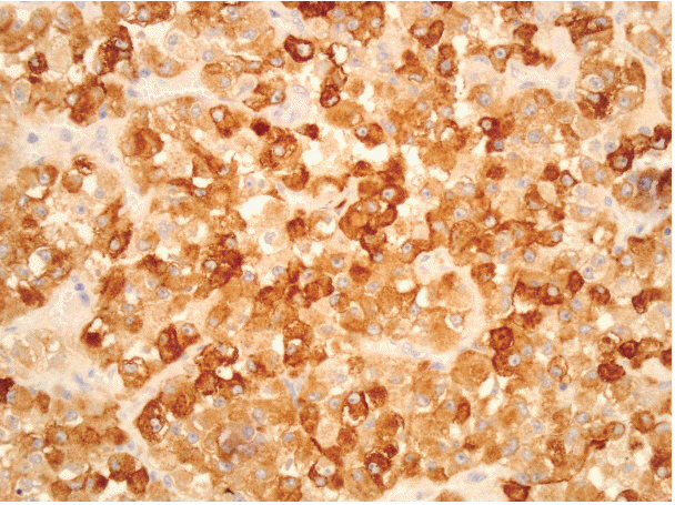
Fig. 2. Human melanoma black 45 immunostaining of the tumor.
Reference
-
1. Kim HY, Choi JH, Lee HS, Choi YJ, Kim A, Kim HK. Sclerosing perivascular epithelioid cell tumor of the lung: a case report with cytologic findings. J Pathol Transl Med. 2016; 50:238–42.
Article2. Cossu A, Paliogiannis P, Tanda F, Dessole S, Palmieri G, Capobianco G. Uterine perivascular epithelioid cell neoplasms (PEComas): report of two cases and literature review. Eur J Gynaecol Oncol. 2014; 35:309–12.3. Goto H, Usui Y, Nagao T. Perivascular epithelioid cell tumor arising from ciliary body treated by local resection. Ocul Oncol Pathol. 2015; 1:88–92.
Article4. Iyengar P, Deangelis DD, Greenberg M, Taylor G. Perivascular epithelioid cell tumor of the orbit: a case report and review of the literature. Pediatr Dev Pathol. 2005; 8:98–104.
Article5. Guthoff R, Guthoff T, Mueller-Hermelink HK, Sold-Darseff J, Geissinger E. Perivascular epithelioid cell tumor of the orbit. Arch Ophthalmol. 2008; 126:1009–11.
Article
- Full Text Links
-
- Actions
-
Cited
- CITED
-
- Close
- Share
-
- Similar articles
-
- A Case of Primary Cutaneous Perivascular Epithelioid Cell Tumor
- Perivascular Epithelioid Cell Tumor in the Stomach
- A Case of Malignant PEComa of the Uterus Associated with Intramural Leiomyoma and Endometrial Carcinoma
- A case of perivascular epithelioid cell tumor (PEComa) at the uterus
- A Case of Malignant Perivascular Epithelioid Cell Tumor of the Retroperitoneum with Multiple Metastases
