A Case of Malignant PEComa of the Uterus Associated with Intramural Leiomyoma and Endometrial Carcinoma
- Affiliations
-
- 1Department of Pathology, Korea University Guro Hospital, Korea University College of Medicine, Seoul, Korea. breakfree83@hanmail.net
- 2Department of Obsterics and Gynecology, Korea University Guro Hospital, Korea University College of Medicine, Seoul, Korea.
- KMID: 2361995
- DOI: http://doi.org/10.4132/jptm.2016.04.20
Abstract
- Perivascular epithelioid cell tumors (PEComas) refers to a family of mesenchymal neoplasms composed of angiomyolipomas, clear cell "sugar" tumors of the lung, and lymphangioleiomyomatoses. These tumors have a distinctive and common component of perivascular epithelioid cells that show an association with blood vessel walls and immunohistochemically display myomelanocytic differentiation. The unique neoplasms have been shown to have an expanded range through a variety of case reports, including visceral, intra-abdominal, soft tissue, and bone tumors. The retroperitoneum, abdominopelvic region, and uterus have been reported to be the most common sites. Most PEComas follow a benign course. However, reports of malignant PEComas are increasing. Many papers have described uterine PEComas, but to our knowledge, there have not yet been any reports of a malignant PEComa arising concomitant with another epithelial tumor and mesenchymal tumor. We report herein the case of a 67-year-old woman who experienced a malignant uterine PEComa infiltrating a preexisting intramural leiomyoma with synchronous well differentiated endometrial carcinoma and multiple liver and lung metastases.
MeSH Terms
Figure
-
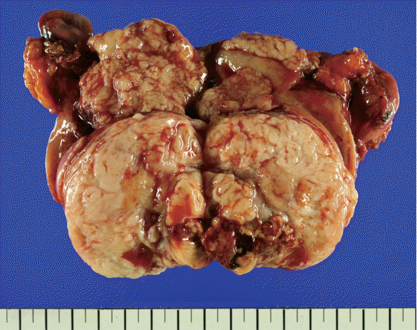
Fig. 1. Total hysterectomy showing endometriod carcinoma on anterior wall to fundus of the endometrium, and whitish leiomyoma encircling perivascular epithelioid cell tumors in the myometrium.

Fig. 2. (A) Endometrioid carcinoma, International Federation of Gynecology and Obstetrics (FIGO) G1. (B) Infiltrating spindle perivascular epithelioid cell tumor (PEComa) cells into surrounding leiomyoma. (C) The PEComa cells have elongated nuclei, prominent nucleoli, clear cytoplasm, cytological atypia, and necrosis.

Fig. 3. (A) Human melanoma black 45 staining shows diffuse cytoplasm and membrane positivity. (B) Transcription factor E3 staining showed nuclear positivity. (C) Desmin stain is negative for perivascular epithelioid cell tumor and positive for leiomyoma. Infiltrative border is identified.
Reference
-
1. Bonetti F, Pea M, Martignoni G, et al. Clear cell (“sugar”) tumor of the lung is a lesion strictly related to angiomyolipoma--the concept of a family of lesions characterized by the presence of the perivascular epithelioid cells (PEC). Pathology. 1994; 26:230–6.2. Fletcher CD, Bridge JA, Hogendoorn P, Mertens F. WHO classification of tumours of soft tissue and bone. 4th ed. Geneva: World Health Organization;2013.3. Bonetti F, Martignoni G, Colato C, et al. Abdominopelvic sarcoma of perivascular epithelioid cells: report of four cases in young women, one with tuberous sclerosis. Mod Pathol. 2001; 14:563–8.
Article4. Folpe AL, Goodman ZD, Ishak KG, et al. Clear cell myomelanocytic tumor of the falciform ligament/ligamentum teres: a novel member of the perivascular epithelioid clear cell family of tumors with a predilection for children and young adults. Am J Surg Pathol. 2000; 24:1239–46.5. Folpe AL, Mentzel T, Lehr HA, Fisher C, Balzer BL, Weiss SW. Perivascular epithelioid cell neoplasms of soft tissue and gynecologic origin: a clinicopathologic study of 26 cases and review of the literature. Am J Surg Pathol. 2005; 29:1558–75.6. Schoolmeester JK, Howitt BE, Hirsch MS, Dal Cin P, Quade BJ, Nucci MR. Perivascular epithelioid cell neoplasm (PEComa) of the gynecologic tract: clinicopathologic and immunohistochemical characterization of 16 cases. Am J Surg Pathol. 2014; 38:176–88.7. Fadare O. Perivascular epithelioid cell tumor (PEComa) of the uterus: an outcome-based clinicopathologic analysis of 41 reported cases. Adv Anat Pathol. 2008; 15:63–75.8. Yu Y, Shi HY, Huang HF. Uterine perivascular epithelioid cell tumour. J Obstet Gynaecol. 2014; 34:519–22.
Article9. Armah HB, Parwani AV. Malignant perivascular epithelioid cell tumor (PEComa) of the uterus with late renal and pulmonary metastases: a case report with review of the literature. Diagn Pathol. 2007; 2:45.
Article10. Argani P, Aulmann S, Illei PB, et al. A distinctive subset of PEComas harbors TFE3 gene fusions. Am J Surg Pathol. 2010; 34:1395–406.
Article11. Malinowska I, Kwiatkowski DJ, Weiss S, Martignoni G, Netto G, Argani P. Perivascular epithelioid cell tumors (PEComas) harboring TFE3 gene rearrangements lack the TSC2 alterations characteristic of conventional PEComas: further evidence for a biological distinction. Am J Surg Pathol. 2012; 36:783–4.12. Gao Z, Bhuiya T, Anderson A. Perivascular epithelioid cell tumour (PEComa) of the uterus associated with malignant neoplasm of the female genital tract. J Obstet Gynaecol. 2004; 24:600–4.
Article13. Folpe AL, Kwiatkowski DJ. Perivascular epithelioid cell neoplasms: pathology and pathogenesis. Hum Pathol. 2010; 41:1–15.
Article
- Full Text Links
-
- Actions
-
Cited
- CITED
-
- Close
- Share
-
- Similar articles
-
- Torsion of a Myomatous Uterus in a Non-Gravid Female: A Case Report
- Endometrial carcinoma arising in a bicornuate uterus
- A Case of Concurrent Uterine Malignant Mixed M llerian Tumor and Endometrial Adenocarcinoma
- Transvaginal Expulsion of Intramural Leiomyoma after Uterine Artery Embolization: Case Report
- A Case of Recurrent Endometrial Carcinoma at the Vagina
