Korean J Gastroenterol.
2014 Nov;64(5):302-306. 10.4166/kjg.2014.64.5.302.
A Case of Malignant Perivascular Epithelioid Cell Tumor of the Retroperitoneum with Multiple Metastases
- Affiliations
-
- 1Department of Internal Medicine, Gyeongsang National University School of Medicine, Jinju, Korea. kimthy@medimail.co.kr
- 2Department of Pathology, Gyeongsang National University School of Medicine, Jinju, Korea.
- 3Institute of Health, Gyeongsang National University School of Medicine, Jinju, Korea.
- KMID: 2164435
- DOI: http://doi.org/10.4166/kjg.2014.64.5.302
Abstract
- Perivascular epithelioid cell tumors (PEComas) are unusual mesenchymal neoplasms composed of histologically and immunohistochemically distinct perivascular epithelioid cells (PECs). Although PEComas have the potential to behave in a malignant fashion, malignant PEComas arising from the retroperitoneum are extremely rare. A 68-year-old woman presented with a painful palpable mass in her left upper abdomen. Computed tomography of the abdomen showed a 9 cm sized heterogeneous mass in left para-aortic space and multiple hypervascular nodules in the liver. 18F-fludeoxyglucose-PET/CT showed multifocal hypermetabolic lesions in retroperitoneum, liver, and skeletal bones. Percutaneous needle biopsies were done on the retroperitoneal and hepatic mass. Both specimens were positive for human melanoma black-45 (HMB-45) on histological and immunohistochemical staining which was compatible with PEComas. Herein, we report a rare case of retroperitoneal PEComa with multiple metastases involving liver and bone at initial diagnosis that exhibited aggressive behavior and resulted in a devastating prognosis.
MeSH Terms
Figure
-

Fig. 1. Abdominal enhanced computer tomography shows multiple hypervascular nodules in the liver (A) and 9 cm sized heterogenous enhancing mass in left para-aortic space (B).
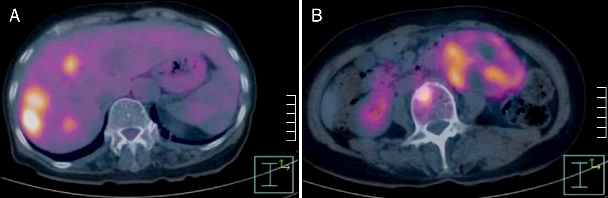
Fig. 2. PET/CT scan shows 18 F-fludeoxyglucose hot uptakes in the liver (A) and left para-aortic space mass (B).
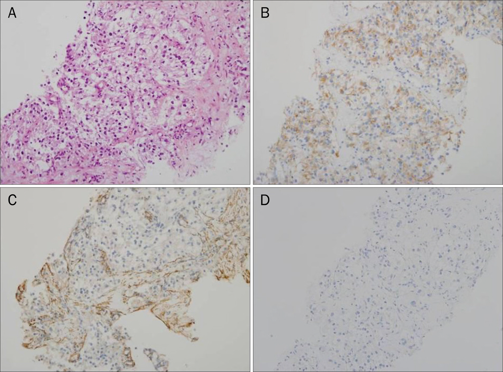
Fig. 3. (A) The tumor shows nested epithelioid tumor cells with round nucleus and granular eosinophilic or clear cytoplasm (H&E, ×200). The tumor cells are positive for HMB-45 (B, ×200), but negative for smooth muscle actin (C, ×200) and S-100 (D, ×200).
Reference
-
References
1. Bonetti F, Pea M, Martignoni G, Zamboni G. PEC and sugar. Am J Surg Pathol. 1992; 16:307–308.
Article2. Pea M, Martignoni G, Zamboni G, Bonetti F. Perivascular epithelioid cell. Am J Surg Pathol. 1996; 20:1149–1153.
Article3. Folpe AL, Kwiatkowski DJ. Perivascular epithelioid cell neoplasms: pathology and pathogenesis. Hum Pathol. 2010; 41:1–15.
Article4. Folpe AL, Mentzel T, Lehr HA, Fisher C, Balzer BL, Weiss SW. Perivascular epithelioid cell neoplasms of soft tissue and gyne-cologic origin: a clinicopathologic study of 26 cases and review of the literature. Am J Surg Pathol. 2005; 29:1558–1575.5. Hornick JL, Fletcher CD. Sclerosing PEComa: clinicopathologic analysis of a distinctive variant with a predilection for the retroperitoneum. Am J Surg Pathol. 2008; 32:493–501.
Article6. Ruco LP, Pilozzi E, Wedard BM, et al. Epithelioid lymphangioleio-myomatosis-like tumour of the uterus in a patient without tuber-ous sclerosis: a lesion mimicking epithelioid leiomyosarcoma. Histopathology. 1998; 33:91–93.
Article7. Hornick JL, Fletcher CD. PEComa: what do we know so far? Histopathology. 2006; 48:75–82.
Article8. Yokoo H, Isoda K, Nakazato Y, et al. Retroperitoneal epithelioid angiomyolipoma leading to fatal outcome. Pathol Int. 2000; 50:649–654.
Article9. Lau SK, Marchevsky AM, McKenna RJ Jr, Luthringer DJ. Malignant monotypic epithelioid angiomyolipoma of the retroperitoneum. Int J Surg Pathol. 2003; 11:223–228.
Article10. Gupta C, Malani AK, Gupta V, Singh J, Ammar H. Metastatic retroperitoneal epithelioid angiomyolipoma. J Clin Pathol. 2007; 60:428–431.
Article11. Lans TE, van Ramshorst GH, Hermans JJ, den Bakker MA, Tran TC, Kazemier G. Perivascular epithelioid cell tumor of the retroperitoneum in a young woman resulting in an abdominal chyloma. J Gastrointest Surg. 2009; 13:389–392.
Article12. Subbiah V, Trent JC, Kurzrock R. Resistance to mammalian tar-get of rapamycin inhibitor therapy in perivascular epithelioid cell tumors. J Clin Oncol. 2010; 28:e415.
Article13. de León DC, Pérez-Montiel D, Bandera A, Villegas C, Gonzalez- Conde E, Vilchis JC. Perivascular epithelioid cell tumor of abdominal origin. Ann Diagn Pathol. 2010; 14:173–177.
Article14. Wagner AJ, Malinowska-Kolodziej I, Morgan JA, et al. Clinical activity of mTOR inhibition with sirolimus in malignant perivascular epithelioid cell tumors: targeting the pathogenic activation of mTORC1 in tumors. J Clin Oncol. 2010; 28:835–840.
Article15. Wu JH, Zhou JL, Cui Y, Jing QP, Shang L, Zhang JZ. Malignant perivascular epithelioid cell tumor of the retroperitoneum. Int J Clin Exp Pathol. 2013; 6:2251–2256.16. Scheppach W, Reissmann N, Sprinz T, Schippers E, Schoettker B, Mueller JG. PEComa of the colon resistant to sirolimus but responsive to doxorubicin/ifosfamide. World J Gastroenterol. 2013; 19:1657–1660.
Article17. Koenig AM, Quaas A, Ries T, et al. Perivascular epitheloid cell tumour (PEComa) of the retroperitoneum - a rare tumor with uncertain malignant behaviour: a case report. J Med Case Rep. 2009; 3:62.
Article18. Gennatas C, Michalaki V, Kairi PV, Kondi-Paphiti A, Voros D. Successful treatment with the mTOR inhibitor everolimus in a patient with perivascular epithelioid cell tumor. World J Surg Oncol. 2012; 10:181.
Article
- Full Text Links
-
- Actions
-
Cited
- CITED
-
- Close
- Share
-
- Similar articles
-
- A Case of Malignant PEComa of the Uterus Associated with Intramural Leiomyoma and Endometrial Carcinoma
- Uncommon of the Uncommon: Malignant Perivascular Epithelioid Cell Tumor of the Lung
- Malignant Perivascular Epithelioid Cell Tumor (PEComa) Arising in the Omentum with Metastatic Peritoneal Seeding: A Case Report
- Primary Perivascular Epithelioid Cell Tumor (PEComa) of the Liver: A Case Report and Review of the Literature
- A Case of Primary Cutaneous Perivascular Epithelioid Cell Tumor
