Combined Effect of Bilateral Ovariectomy and Anterior Cruciate Ligament Transection With Medial Meniscectomy on the Development of Osteoarthritis Model
- Affiliations
-
- 1Department of Physical and Rehabilitation Medicine, Samsung Medical Center, Sungkyunkwan University School of Medicine, Seoul, Korea. guitarren.kim@samsung.com
- KMID: 2356643
- DOI: http://doi.org/10.5535/arm.2016.40.4.583
Abstract
OBJECTIVE
To investigate the combined effect of bilateral ovariectomy (OVX) and anterior cruciate ligament transection (ACLT) with medial meniscectomy (MM) on the development of osteoarthritis (OA).
METHODS
Twenty female 15-week-old Sprague-Dawley rats were used. Five rats in each group underwent bilateral OVX (OVX group), bilateral ACLT with MM (ACLT with MM group), bilateral OVX plus ACLT with MM (OVX plus ACLT with MM group), and sham surgery (SHAM group). All the rats were subjected to treadmill running for 4 weeks. The behavioral evaluation for induction of OA used the number of rears method, and this was conducted at 1, 2, and 4 weeks post-surgery. Bone mineral density (BMD) was calculated with micro-computerized tomography images and the modified Mankin's scoring was used for the histological changes.
RESULTS
The number of rears in the OVX plus ACLT with MM group decreased gradually and more rapidly in the ACLT with MM group. Histologically, the OVX plus ACLT with MM group had a significantly higher modified Mankin's score than the OVX group (p=0.008) and the SHAM group (p=0.008). BMDs of the OVX plus ACLT with MM group were significantly lower than the SHAM group (p=0.002), and the ACLT with MM group (p=0.003).
CONCLUSION
We found that bilateral OVX plus ACLT with MM induced definite OA change in terms of histology and BMD compared to bilateral OVX and ACLT with MM alone. Therefore, OVX and ACLT with MM was an appropriate degenerative OA rat model.
MeSH Terms
Figure
-
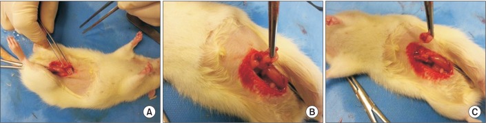
Fig. 1 Details shown for the ovariectomy procedure. (A) A 2- to 3-cm ventral midline skin incision and a 2-cm midline muscle incision were made at the level of uterus. (B) After the ligation of the ovarian duct, (C) the ovary was extracted.
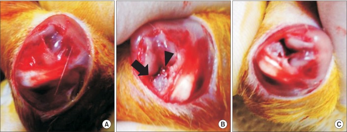
Fig. 2 (A) A 4-cm vertical midline incision was made in the skin and fascia on the knee cap region of the left hind limb. (B) The patella was pushed laterally and the synovial membrane was excised and the knee joint was bent to expose the anterior cruciate ligament (solid triangle) and the medial meniscus (solid arrow). (C) The anterior cruciate ligament was transected and the medial meniscus was completely removed.
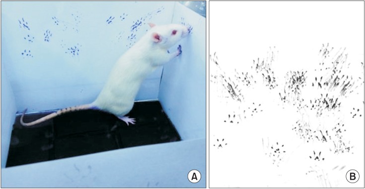
Fig. 3 Measurement of the number of rears. (A) With paws covered in foam stamp pad, the rats stood on their hind limbs and touched the walls of the box with their forelimbs. (B) The footprints left on the paper.
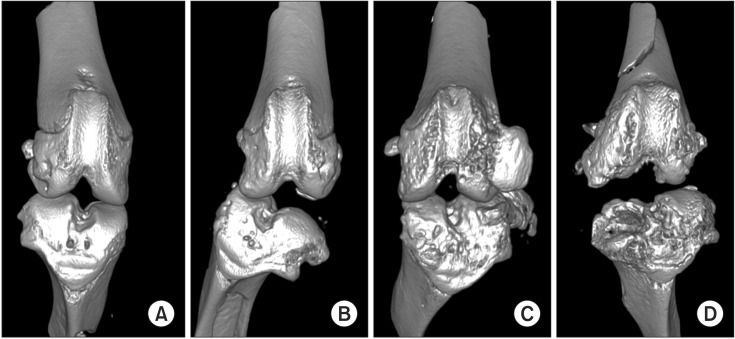
Fig. 4 Micro-computed tomography images of knee joints with three-dimensional reconstruction. Each image is from a different surgery group. (A) Sham surgery, (B) bilateral ovariectomy, (C) anterior cruciate ligament transection with medial meniscectomy, and (D) bilateral ovariectomy and anterior cruciate ligament transection with medial meniscectomy.
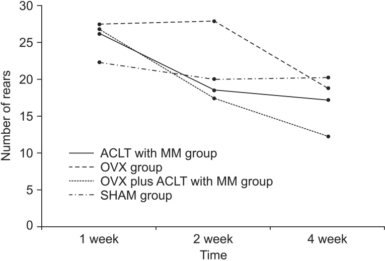
Fig. 5 The number of rears at 1, 2, and 4 weeks after surgery in the sham surgery group (SHAM group), the bilateral ovariectomy group (OVX group), the anterior cruciate ligament transection with medial meniscectomy group (ACLT with MM group), and the bilateral ovariectomy plus anterior cruciate ligament transection with medial meniscectomy group (OVX plus ACLT with MM group). This figure shows significant decrease in the number of rears for the overall groups (p<0.05 for time effect), but there was no significant difference between the groups in the number of rears (p=0.542).
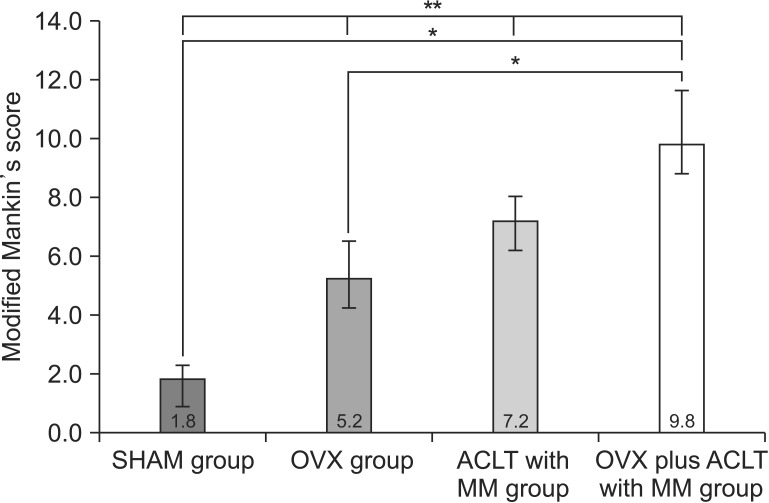
Fig. 6 Modified Mankin's scores of the sham surgery group (SHAM group), the bilateral ovariectomy group (OVX group), the anterior cruciate ligament transection with medial meniscectomy group (ACLT with MM group), and the bilateral ovariectomy plus anterior cruciate ligament transection with medial meniscectomy group (OVX plus ACLT with MM group). This figure shows significant differences among all groups (**p<0.001). OVX plus ACLT with MM group showed significantly higher modified Mankin's score than the OVX group (*p=0.008) and the SHAM group (*p=0.008). The Kruskal-Wallis test followed by post-hoc Mann-Whitney U test with Bonferroni correction.
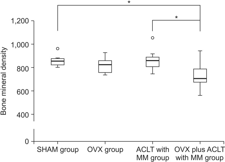
Fig. 7 Bone mineral density of the sham surgery group (SHAM group), the bilateral ovariectomy group (OVX group), the anterior cruciate ligament transection with medial meniscectomy group (ACLT with MM group), and the bilateral ovariectomy plus anterior cruciate ligament transection with medial meniscectomy group (OVX plus ACLT with MM group). There was a significant difference of bone mineral densities between not only the SHAM group and the OVX plus ACLT with MM group (*p=0.003) but also the ACLT with MM group and the OVX plus ACLT with MM group (*p=0.002). Small circle (○) is outlier. The Kruskal-Wallis test followed by post-hoc Mann-Whitney U test with Bonferroni correction.
Reference
-
1. Spector TD, Hart DJ, Byrne J, Harris PA, Dacre JE, Doyle DV. Definition of osteoarthritis of the knee for epidemiological studies. Ann Rheum Dis. 1993; 52:790–794. PMID: 8250610.
Article2. Little CB, Hunter DJ. Post-traumatic osteoarthritis: from mouse models to clinical trials. Nat Rev Rheumatol. 2013; 9:485–497. PMID: 23689231.
Article3. Poole R, Blake S, Buschmann M, Goldring S, Laverty S, Lockwood S, et al. Recommendations for the use of preclinical models in the study and treatment of osteoarthritis. Osteoarthritis Cartilage. 2010; 18 Suppl 3:S10–S16. PMID: 20864015.
Article4. Pickarski M, Hayami T, Zhuo Y, Duong LT. Molecular changes in articular cartilage and subchondral bone in the rat anterior cruciate ligament transection and meniscectomized models of osteoarthritis. BMC Musculoskelet Disord. 2011; 12:197. PMID: 21864409.
Article5. Sniekers YH, Weinans H, Bierma-Zeinstra SM, van Leeuwen JP, van Osch GJ. Animal models for osteoarthritis: the effect of ovariectomy and estrogen treatment. A systematic approach. Osteoarthritis Cartilage. 2008; 16:533–541. PMID: 18280756.6. Zhang Y, McAlindon TE, Hannan MT, Chaisson CE, Klein R, Wilson PW, et al. Estrogen replacement therapy and worsening of radiographic knee osteoarthritis: the Framingham Study. Arthritis Rheum. 1998; 41:1867–1873. PMID: 9778229.
Article7. Turner AS, Athanasiou KA, Zhu CF, Alvis MR, Bryant HU. Biochemical effects of estrogen on articular cartilage in ovariectomized sheep. Osteoarthritis Cartilage. 1997; 5:63–69. PMID: 9010879.
Article8. Reis EM, Ropke J, Busanello A, Reckziegel P, Leal CQ, Wagner C, et al. Effect of Hypericum perforatum on different models of movement disorders in rats. Behav Pharmacol. 2013; 24:623–627. PMID: 23962987.
Article9. Fowler SC, Muma NA. Use of a force-sensing automated open field apparatus in a longitudinal study of multiple behavioral deficits in CAG140 Huntington's disease model mice. Behav Brain Res. 2015; 294:7–16. PMID: 26210937.
Article10. Nagase H, Kumakura S, Shimada K. Establishment of a novel objective and quantitative method to assess pain-related behavior in monosodium iodoacetate-induced osteoarthritis in rat knee. J Pharmacol Toxicol Methods. 2012; 65:29–36. PMID: 22037051.
Article11. Kuroki H, Nakagawa Y, Mori K, Ohba M, Suzuki T, Mizuno Y, et al. Acoustic stiffness and change in plug cartilage over time after autologous osteochondral grafting: correlation between ultrasound signal intensity and histological score in a rabbit model. Arthritis Res Ther. 2004; 6:R492–R504. PMID: 15535827.12. Ostergaard K, Andersen CB, Petersen J, Bendtzen K, Salter DM. Validity of histopathological grading of articular cartilage from osteoarthritic knee joints. Ann Rheum Dis. 1999; 58:208–213. PMID: 10364898.
Article13. Pauli C, Whiteside R, Heras FL, Nesic D, Koziol J, Grogan SP, et al. Comparison of cartilage histopathology assessment systems on human knee joints at all stages of osteoarthritis development. Osteoarthritis Cartilage. 2012; 20:476–485. PMID: 22353747.
Article14. Chan WP, Lang P, Stevens MP, Sack K, Majumdar S, Stoller DW, et al. Osteoarthritis of the knee: comparison of radiography, CT, and MR imaging to assess extent and severity. AJR Am J Roentgenol. 1991; 157:799–806. PMID: 1892040.
Article15. Ferrandiz ML, Terencio MC, Carceller MC, Ruhí R, Dalmau P, Verges J, et al. Effects of BIS076 in a model of osteoarthritis induced by anterior cruciate ligament transection in ovariectomised rats. BMC Musculoskelet Disord. 2015; 16:92. PMID: 25903377.
Article16. Hart DA, Achari Y. Alterations to cell metabolism in connective tissues of the knee after ovariohysterectomy in a rabbit model: are there implications for the postmenopausal athlete. Br J Sports Med. 2010; 44:867–871. PMID: 19136500.
Article17. Yoshida A, Morihara T, Matsuda K, Sakamoto H, Arai Y, Kida Y, et al. Immunohistochemical analysis of the effects of estrogen on intraarticular neurogenic inflammation in a rat anterior cruciate ligament transection model of osteoarthritis. Connect Tissue Res. 2012; 53:197–206. PMID: 22141435.
Article18. Finkelstein JS, Brockwell SE, Mehta V, Greendale GA, Sowers MR, Ettinger B, et al. Bone mineral density changes during the menopause transition in a multiethnic cohort of women. J Clin Endocrinol Metab. 2008; 93:861–868. PMID: 18160467.
Article19. Lindsay R, Hart DM, Sweeney A, Coutts JR, Clarke A. Endogenous oestrogen and bone loss following oophorectomy. Calcif Tissue Res. 1977; 22 Suppl:213–216. PMID: 912525.
Article20. Martin-Millan M, Castaneda S. Estrogens, osteoarthritis and inflammation. Joint Bone Spine. 2013; 80:368–373. PMID: 23352515.
Article21. Im GI, Kim MK. The relationship between osteoarthritis and osteoporosis. J Bone Miner Metab. 2014; 32:101–109. PMID: 24196872.
Article22. Hannan MT, Anderson JJ, Zhang Y, Levy D, Felson DT. Bone mineral density and knee osteoarthritis in elderly men and women: the Framingham study. Arthritis Rheum. 1993; 36:1671–1680. PMID: 8250986.
Article23. Karvonen RL, Miller PR, Nelson DA, Granda JL, Fernandez-Madrid F. Periarticular osteoporosis in osteoarthritis of the knee. J Rheumatol. 1998; 25:2187–2194. PMID: 9818663.24. Lee JY, Harvey WF, Price LL, Paulus JK, Dawson-Hughes B, McAlindon TE. Relationship of bone mineral density to progression of knee osteoarthritis. Arthritis Rheum. 2013; 65:1541–1546. PMID: 23494470.
Article25. Inada M, Matsumoto C, Miyaura C. Animal models for bone and joint disease: ovariectomized and orchidectomized animals. Clin Calcium. 2011; 21:164–170. PMID: 21289412.
- Full Text Links
-
- Actions
-
Cited
- CITED
-
- Close
- Share
-
- Similar articles
-
- Results of Anterior Cruciate Ligament Reconstruction with Unicondylar Arthroplasty for Medial Compartment Knee Osteoarthritis combined with Anterior Instability
- Anomalous Insertion of the Anterior Medial Meniscus
- The Change of the Mechanoreceptor fo Anterior Cruciate Ligament after Injuries of Medial Articular Cartilage and Ligament in the Rabbit
- A Study on the Development of Degenerative Osteoarthritis after Arthroscopic Total Menisectomy
- Pitalls in Interpretation of Physical Tests of Knee Ligament Injury
