J Cardiovasc Ultrasound.
2016 Sep;24(3):208-214. 10.4250/jcu.2016.24.3.208.
Diastolic Dyssynchrony in Acute ST Segment Elevation Myocardial Infarction and Relationship with Functional Recovery of Left Ventricle
- Affiliations
-
- 1Cardiology Department, Kocaeli Derince Training and Research Hospital, Kocaeli, Turkey. drburakturan@gmail.com
- 2Cardiology Department, School of Medicine, Marmara University, Istanbul, Turkey.
- KMID: 2353098
- DOI: http://doi.org/10.4250/jcu.2016.24.3.208
Abstract
- BACKGROUND
Incidence of diastolic dyssynchrony (DD) and its impact on functional recovery of left ventricle (LV) after ST segment elevation myocardial infarction (STEMI) is not known.
METHODS
Consecutive patients with STEMI who underwent successful revascularization were prospectively enrolled. Echocardiography with tissue Doppler imaging was performed within 48 hours of admission and at 6 months. LV end-diastolic volume index (EDVI), end-systolic volume index (ESVI), ejection fraction (EF), and left atrial volume index (LAVI) were calculated. Diastolic delay was calculated from onset of QRS complex to peak of E wave in tissue Doppler image and presented as maximal temporal difference between peak early diastolic velocity of 6 basal segments of LV (TeDiff). Study patients were compared with demographically matched control group.
RESULTS
Forty eight consecutive patients (55 ± 10 years, 88% male) and 24 controls (56 ± 6 years, 88% male) were included. TeDiff was higher in STEMI than in controls (35.9 ± 19.9 ms vs. 26.3 ± 6.8 ms, p = 0.025). Presence of DD was higher in STEMI than controls (58% vs. 33%, p = 0.046) according to calculated cut-off value (≥ 29 ms). There was no correlation between TeDiff and change in EDVI, ESVI, and LAVI at 6 months, however TeDiff and change in EF at 6 months was positively correlated (r = 0.328, p = 0.023). Patients with baseline DD experienced remodeling less frequently compared to patients without baseline DD (11% vs. 38%, p = 0.040) during follow-up.
CONCLUSION
STEMI disrupts diastolic synchronicity of LV. However, DD during acute phase of STEMI is associated with better recovery of LV thereafter. This suggests that DD is associated with peri-infarct stunned myocardium that is salvaged with primary intervention as well as infarct size.
MeSH Terms
Figure
-

Fig. 1 Maximal diastolic delay between 6 basal segments of LV (TeDiff) of controls, patients during and 6 months after STEMI. Bars indicate means and standard errors. LV: left ventricle, STEMI: ST segment elevation myocardial infarction.
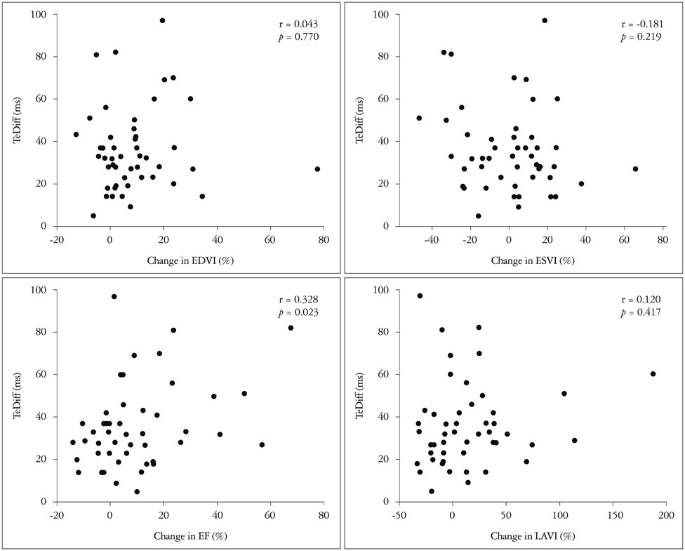
Fig. 2 Correlation of TeDiff with % change in EDVI, ESVI, EF, and LAVI. TeDiff: maximal temporal difference between peak early diastolic velocity of 6 basal segments, EDVI: end-diastolic volume index, ESVI: end-systolic volume index, EF: ejection fraction, LAVI: left atrial volume index.
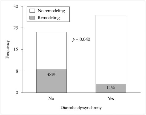
Fig. 3 Incidence of late remodeling according to presence of baseline diastolic dyssynchrony (TeDiff ≥ 29 ms). TeDiff: maximal temporal difference between peak early diastolic velocity of 6 basal segments.
Reference
-
1. Prasad SB, See V, Tan T, Brown P, McKay T, Kovoor P, Thomas L. Serial Doppler echocardiographic assessment of diastolic dysfunction during acute myocardial infarction. Echocardiography. 2012; 29:1164–1171.2. Bonow RO, Bacharach SL, Green MV, Kent KM, Rosing DR, Lipson LC, Leon MB, Epstein SE. Impaired left ventricular diastolic filling in patients with coronary artery disease: assessment with radionuclide angiography. Circulation. 1981; 64:315–323.3. Temporelli PL, Giannuzzi P, Nicolosi GL, Latini R, Franzosi MG, Gentile F, Tavazzi L, Maggioni AP. GISSI-3 Echo Substudy Investigators. Doppler-derived mitral deceleration time as a strong prognostic marker of left ventricular remodeling and survival after acute myocardial infarction: results of the GISSI-3 echo substudy. J Am Coll Cardiol. 2004; 43:1646–1653.4. Ueno Y, Nakamura Y, Kinoshita M, Fujita T, Sakamoto T, Okamura H. An early predictor of left ventricular remodeling after reperfused anterior acute myocardial infarction: ratio of peak E wave velocity/flow propagation velocity and mitral E wave deceleration time. Echocardiography. 2002; 19(7 Pt 1):555–563.5. Nucifora G, Bertini M, Marsan NA, Delgado V, Scholte AJ, Ng AC, van Werkhoven JM, Siebelink HM, Holman ER, Schalij MJ, van der Wall EE, Bax JJ. Impact of left ventricular dyssynchrony early on left ventricular function after first acute myocardial infarction. Am J Cardiol. 2010; 105:306–311.6. Turan B, Yilmaz F, Karaahmet T, Tigen K, Mutlu B, Basaran Y. Role of left ventricular dyssynchrony in predicting remodeling after ST elevation myocardial infarction. Echocardiography. 2012; 29:165–172.7. Grines CL, Bashore TM, Boudoulas H, Olson S, Shafer P, Wooley CF. Functional abnormalities in isolated left bundle branch block. The effect of interventricular asynchrony. Circulation. 1989; 79:845–853.8. Chang SA, Kim HK, Kim DH, Kim YJ, Sohn DW, Oh BH, Park YB. Left ventricular systolic and diastolic dyssynchrony in asymptomatic hypertensive patients. J Am Soc Echocardiogr. 2009; 22:337–342.9. Ko JS, Jeong MH, Lee MG, Lee SE, Kang WY, Kim SH, Park KH, Sim DS, Yoon NS, Yoon HJ, Hong YJ, Park HW, Kim JH, Ahn Y, Cho JG, Park JC, Kang JC. Left ventricular dyssynchrony after acute myocardial infarction is a powerful indicator of left ventricular remodeling. Korean Circ J. 2009; 39:236–242.10. Kang SJ, Song JK, Yang HS, Song JM, Kang DH, Rhee KS, Nam GB, Choi KJ, Kim JJ, Kim YH. Systolic and diastolic regional myocardial motion of pacing-induced versus idiopathic left bundle branch block with and without left ventricular dysfunction. Am J Cardiol. 2004; 93:1243–1246.11. Yu CM, Lin H, Zhang Q, Sanderson JE. High prevalence of left ventricular systolic and diastolic asynchrony in patients with congestive heart failure and normal QRS duration. Heart. 2003; 89:54–60.12. Schuster I, Habib G, Jego C, Thuny F, Avierinos JF, Derumeaux G, Beck L, Medail C, Franceschi F, Renard S, Ferracci A, Lefevre J, Luccioni R, Deharo JC, Djiane P. Diastolic asynchrony is more frequent than systolic asynchrony in dilated cardiomyopathy and is less improved by cardiac resynchronization therapy. J Am Coll Cardiol. 2005; 46:2250–2257.13. Shanks M, Bertini M, Delgado V, Ng AC, Nucifora G, van Bommel RJ, Borleffs CJ, Holman ER, van de Veire NR, Schalij MJ, Bax JJ. Effect of biventricular pacing on diastolic dyssynchrony. J Am Coll Cardiol. 2010; 56:1567–1575.14. Yu CM, Zhang Q, Yip GW, Lee PW, Kum LC, Lam YY, Fung JW. Diastolic and systolic asynchrony in patients with diastolic heart failure: a common but ignored condition. J Am Coll Cardiol. 2007; 49:97–105.15. Sun JP, Xu TY, Lee AP, Yang XS, Liu M, Li Y, Wang JG, Yu CM. Early diastolic dyssynchrony in relation to left ventricular remodeling and function in hypertension. Int J Cardiol. 2015; 179:195–200.16. Lee PW, Zhang Q, Yip GW, Wu L, Lam YY, Wu EB, Yu CM. Left ventricular systolic and diastolic dyssynchrony in coronary artery disease with preserved ejection fraction. Clin Sci (Lond). 2009; 116:521–529.17. Bonow RO, Vitale DF, Bacharach SL, Frederick TM, Kent KM, Green MV. Asynchronous left ventricular regional function and impaired global diastolic filling in patients with coronary artery disease: reversal after coronary angioplasty. Circulation. 1985; 71:297–307.18. Perrone-Filardi P, Bacharach SL, Dilsizian V, Bonow RO. Effects of regional systolic asynchrony on left ventricular global diastolic function in patients with coronary artery disease. J Am Coll Cardiol. 1992; 19:739–744.19. Mollema SA, Liem SS, Suffoletto MS, Bleeker GB, van der Hoeven BL, van de Veire NR, Boersma E, Holman ER, van der Wall EE, Schalij MJ, Gorcsan J 3rd, Bax JJ. Left ventricular dyssynchrony acutely after myocardial infarction predicts left ventricular remodeling. J Am Coll Cardiol. 2007; 50:1532–1540.20. Chang SA, Chang HJ, Choi SI, Chun EJ, Yoon YE, Kim HK, Kim YJ, Choi DJ, Sohn DW, Helm RH, Lardo AC. Usefulness of left ventricular dyssynchrony after acute myocardial infarction, assessed by a tagging magnetic resonance image derived metric, as a determinant of ventricular remodeling. Am J Cardiol. 2009; 104:19–23.21. Zhang Y, Yip GW, Chan AK, Wang M, Lam WW, Fung JW, Chan JY, Sanderson JE, Yu CM. Left ventricular systolic dyssynchrony is a predictor of cardiac remodeling after myocardial infarction. Am Heart J. 2008; 156:1124–1132.
- Full Text Links
-
- Actions
-
Cited
- CITED
-
- Close
- Share
-
- Similar articles
-
- ST segment
- Differences in Clinical Outcomes Between Patients With ST-Elevation Versus Non-ST-Elevation Acute Myocardial Infarction in Korea
- Precordial ST-Segment Elevation in Acute Right Ventricular Myocardial Infarction
- The New Diagnostic Algorithm for New or Presumably New Left Bundle Branch Block and Suspected Acute Myocardial Infarction
- The Eletrocardiographic Analysis of Acute Myocardial Infarction and Non-infarction Syndrome In the Patients with ST Segment Elevation and Chest Pain
