Attenuation of Peripheral Regulatory T-Cell Suppression of Skin-Homing CD8+T Cells in Atopic Dermatitis
- Affiliations
-
- 1Department of Dermatology, Yidu Central Hospital, Weifang Medical University, Weifang, P.R. China.
- 2Department of Health Statistics, Public Health College of Weifang Medical University, Weifang, P.R. China.
- 3Department of Clinical Laboratory, Yidu Central Hospital, Weifang Medical University, Weifang, P.R. China.
- 4Department of Dermatology, Weifang Skin Disease Hospital, Weifang, P.R. China.
- 5Department of Stomatology, Yidu Central Hospital, Weifang Medical University, Weifang, P.R. China. dentistdg@sina.com
- KMID: 2352807
- DOI: http://doi.org/10.3349/ymj.2015.56.1.196
Abstract
- PURPOSE
Cutaneous lymphocyte-associated antigen (CLA)-expressing CD8+T cells have been known to play an important role in the pathogenesis of atopic dermatitis (AD). However, the mechanisms underlying the loss of self-tolerance remain unclear. Regulatory T cells (Tregs) play a key role in the development of homeostasis in the immune system. We, therefore, hypothesized that a reduced ability of Tregs to inhibit autologous CD8+CLA+T cells might be underlying mechanism in AD.
MATERIALS AND METHODS
CD8+CLA+T cells and Tregs were obtained from the peripheral blood of AD patients and control volunteers. The frequencies of CD8+CLA+T cells were evaluated. The proliferative responses of CD8+CLA+T cells were assessed by flow cytometry, and the levels of transforming growth factor-beta1 (TGF-beta1) and interleukin-10 (IL-10) in culture supernatants were detected by enzyme-linked immunosorbent assay.
RESULTS
Our results revealed higher frequency and increased expression of perforin and granzyme-B in peripheral CD8+CLA+T cells in AD, and lower inhibitory ability of Tregs on proliferation of CD8+CLA+T cells in AD. Meanwhile, the levels of TGF-beta1 produced by Tregs were significantly lower in AD, and anti-TGF-beta1 abolished such suppression.
CONCLUSION
The attenuated inhibitory ability of Tregs on hyper-activated autologous CD8+CLA+T cells, mediated by TGF-beta1, plays an important role in the pathogenesis of AD.
MeSH Terms
-
Adult
Aged
CD8-Positive T-Lymphocytes/drug effects/*immunology
Case-Control Studies
Cell Proliferation
Cell Separation
Dermatitis, Atopic/*immunology/pathology
Female
Granzymes/metabolism
Humans
Interleukin-10/metabolism
Lymphocyte Count
Male
Perforin/metabolism
Skin/*immunology/pathology
T-Lymphocytes, Cytotoxic/drug effects/immunology
T-Lymphocytes, Regulatory/drug effects/*immunology
Transforming Growth Factor beta1/pharmacology
Granzymes
Interleukin-10
Perforin
Transforming Growth Factor beta1
Figure
-
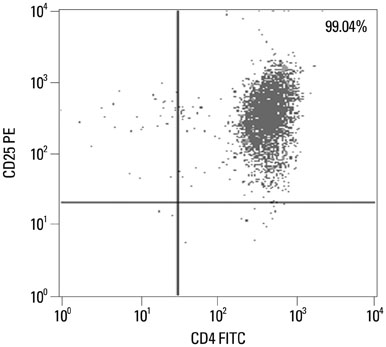
Fig. 1 The purities of isolated Tregs (CD4+CD25+T cells). PBMCs were isolated on Ficoll-Hypaque gradient. According to the manufacturer's instructions of CD4+CD25+ Regulatory T Cell Isolation Kit, CD4+T cells were purified by negtive selection, and then CD25+T cells were isolated by positive selection. The purities were confirmed >95%. A representative profile of purified Tregs was showed. PBMC, peripheral blood mononuclear cell; FITC, fluorescein isothiocyanate; PE, phycoerythrin.

Fig. 2 Frequencies of peripheral CD8+CLA+T cells. The frequencies of CD8+CLA+T cells in peripheral blood were measured by flow cytometry, and the representative results (A) from AD patients and healthy volunteers were shown by dot plot graphs. (B) The data showed the frequencies of CD8+CLA+T cells were significantly higher in the AD group (n=30) compared to the control group (n=25, *p<0.05). CLA, cutaneous lymphocyte-associated antigen; AD, atopic dermatitis; PE, phycoerythrin.

Fig. 3 Expression levels of cytotoxic molecules (perforin and granzyme-B) on peripheral CD8+CLA+T cells. The percentages of cytotoxic molecules (perforin and granzyme-B) on CD8+CLA+T cells in peripheral blood were measured by flow cytometry, and the representative results (A and C) from AD patients and healthy volunteers were shown by dot plot graphs. (B and D) The data showed the expression levels of both perforin and granzyme-B on CD8+CLA+T cells were significantly higher in the AD group (n=30) compared to the control group (n=25, *p<0.05). CLA, cutaneous lymphocyte-associated antigen; AD, atopic dermatitis; PE, phycoerythrin; FITC, fluorescein isothiocyanate.
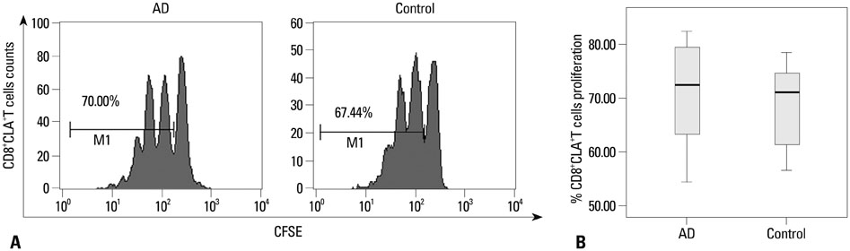
Fig. 4 Proliferation of peripheral CD8+CLA+T cells. CFSE-labeled peripheral CD8+CLA+T cells stimulated with anti-CD3/CD28 in the absence of Tregs for 4 days. According to the CFSE intensity by flow cytometry, the percentages of proliferating CD8+CLA+T cells were detected. (A) Representative CFSE profiles from an AD patient and a healthy volunteer were shown. (B) The data showed the percentages of proliferating CD8+CLA+T cells were of no significant difference between the AD group (n=8) and the control group (n=8). CFSE, carboxyfluorescein diacetate succinimidyl ester; CLA, cutaneous lymphocyte-associated antigen; AD, atopic dermatitis.
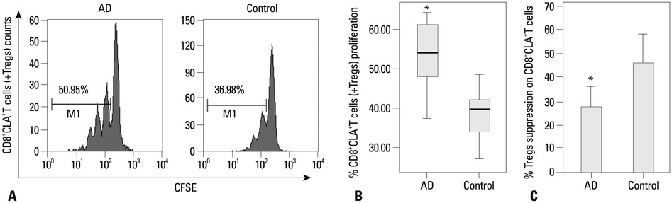
Fig. 5 Tregs suppress proliferation of autologous CD8+CLA+T cells. CFSE-labeled peripheral CD8+CLA+T cells stimulated with anti-CD3/CD28 in the presence of autologous Tregs for 4 days. According to the CFSE intensity by flow cytometry, the percentages of proliferating CD8+CLA+T cells were detected. (A) Representative CFSE profiles from an AD patient and a healthy volunteer were shown. (B) The data showed the percentages of proliferating CD8+CLA+T cells were significantly higher in the AD group (n=8) compared to the control group (n=8, *p<0.05). (C) The suppressive function of Tregs on the proliferation of autologous CD8+CLA+T cells was significantly lower in the AD group (n=8) compared to the control group (n=8, *p<0.05). CFSE, carboxyfluorescein diacetate succinimidyl ester; Tregs, regulatory T cells; CLA, cutaneous lymphocyte-associated antigen; AD, atopic dermatitis.
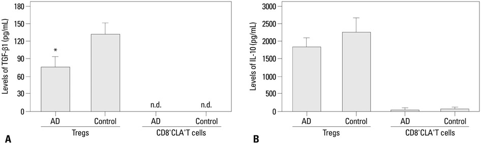
Fig. 6 Levels of TGF-β1 and IL-10 in culture supernatants of Tregs and/or CD8+CLA+T cells. 5×10⁴ Tregs co-cultured with 5×10⁴ autologous CD8+CLA+T cells stimulated with anti-CD3/CD28 in 96-well plates. Four days later, the amount of TGF-β1 (A) and IL-10 (B) in culture supernatant was measured by ELISA. The levels of TGF-β1 produced by Tregs were significantly lower in the AD group (n=8) compared to the control group (n=8, *p<0.05). However, the levels of IL-10 produced by Tregs showed no difference between the two groups. And 5×10⁴ CD8+CLA+T cells stimulated with anti-CD3/CD28 in 96-well plates for 4 days, secreted undetectable amounts of TGF-β1 and minimal amounts of IL-10 in the two groups. CLA, cutaneous lymphocyte-associated antigen; AD, atopic dermatitis; Tregs, regulatory T cells; ELISA, enzyme-linked immunosorbent assay; TGF-β1, transforming growth factor-β1; IL-10, interleukin-10.
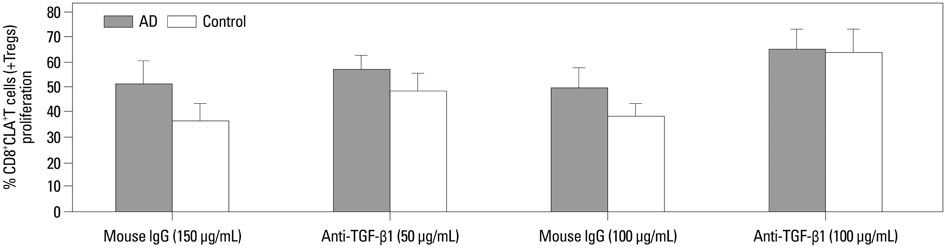
Fig. 7 Suppression of CD8+CLA+T cells proliferation by Tregs is mediated by TGF-β1. 5×10⁴ Tregs co-cultured with 5×10⁴ autologous CD8+CLA+T cells from AD patients (n=8, gray bars) or healthy volunteers (n=8, white bars) stimulated with anti-CD3/CD28 in 96-well plates in the presence of anti-TGF-β1 or control IgG. Four days later, the percentages of proliferating CD8+CLA+T cells were detected. The CD8+CLA+T cells exhibited increased proliferation in the presence of 50 µg/mL of anti-TGF-β1, and their proliferation were further increased and of no significant difference between the two groups in the presence of 100 µg/mL of anti-TGF-β1. Tregs, regulatory T cells; CLA, cutaneous lymphocyte-associated antigen; AD, atopic dermatitis; TGF-β1, transforming growth factor-β1.
Reference
-
1. Bieber T. Atopic dermatitis. N Engl J Med. 2008; 358:1483–1494.
Article2. Hennino A, Jean-Decoster C, Giordano-Labadie F, Debeer S, Vanbervliet B, Rozières A, et al. CD8+ T cells are recruited early to allergen exposure sites in atopy patch test reactions in human atopic dermatitis. J Allergy Clin Immunol. 2011; 127:1064–1067.
Article3. Hennino A, Vocanson M, Toussaint Y, Rodet K, Benetière J, Schmitt AM, et al. Skin-infiltrating CD8+ T cells initiate atopic dermatitis lesions. J Immunol. 2007; 178:5571–5577.
Article4. Sallusto F, Lenig D, Förster R, Lipp M, Lanzavecchia A. Two subsets of memory T lymphocytes with distinct homing potentials and effector functions. Nature. 1999; 401:708–712.
Article5. Akdis M, Simon HU, Weigl L, Kreyden O, Blaser K, Akdis CA. Skin homing (cutaneous lymphocyte-associated antigen-positive) CD8+ T cells respond to superantigen and contribute to eosinophilia and IgE production in atopic dermatitis. J Immunol. 1999; 163:466–475.6. Teraki Y, Hotta T, Shiohara T. Increased circulating skin-homing cutaneous lymphocyte-associated antigen (CLA)+ type 2 cytokine-producing cells, and decreased CLA+ type 1 cytokine-producing cells in atopic dermatitis. Br J Dermatol. 2000; 143:373–378.
Article7. Sakaguchi S. Naturally arising CD4+ regulatory t cells for immunologic self-tolerance and negative control of immune responses. Annu Rev Immunol. 2004; 22:531–562.8. Loser K, Hansen W, Apelt J, Balkow S, Buer J, Beissert S. In vitro-generated regulatory T cells induced by Foxp3-retrovirus infection control murine contact allergy and systemic autoimmunity. Gene Ther. 2005; 12:1294–1304.
Article9. Fontenot JD, Gavin MA, Rudensky AY. Foxp3 programs the development and function of CD4+CD25+ regulatory T cells. Nat Immunol. 2003; 4:330–336.
Article10. Hori S, Nomura T, Sakaguchi S. Control of regulatory T cell development by the transcription factor Foxp3. Science. 2003; 299:1057–1061.
Article11. Verhagen J, Akdis M, Traidl-Hoffmann C, Schmid-Grendelmeier P, Hijnen D, Knol EF, et al. Absence of T-regulatory cell expression and function in atopic dermatitis skin. J Allergy Clin Immunol. 2006; 117:176–183.
Article12. Caproni M, Antiga E, Torchia D, Volpi W, Barletta E, Gitti G, et al. FoxP3-expressing T regulatory cells in atopic dermatitis lesions. Allergy Asthma Proc. 2007; 28:525–528.
Article13. Schnopp C, Rad R, Weidinger A, Weidinger S, Ring J, Eberlein B, et al. Fox-P3-positive regulatory T cells are present in the skin of generalized atopic eczema patients and are not particularly affected by medium-dose UVA1 therapy. Photodermatol Photoimmunol Photomed. 2007; 23:81–85.
Article14. Szegedi A, Baráth S, Nagy G, Szodoray P, Gál M, Sipka S, et al. Regulatory T cells in atopic dermatitis: epidermal dendritic cell clusters may contribute to their local expansion. Br J Dermatol. 2009; 160:984–993.
Article15. Hanifin JM, Rajka G. Diagnostic features of atopic dermatitis. Acta Derm Venereol (Stockh 92). 1980; Suppl 92. 44–47.16. Sigmundsdóttir H, Gudjónsson JE, Jónsdóttir I, Lúdvíksson BR, Valdimarsson H. The frequency of CLA+ CD8+ T cells in the blood of psoriasis patients correlates closely with the severity of their disease. Clin Exp Immunol. 2001; 126:365–369.
Article17. Yano S, Nakamura K, Okochi H, Tamaki K. Analysis of the expression of cutaneous lymphocyte-associated antigen on the peripheral blood and cutaneous lymphocytes of alopecia areata patients. Acta Derm Venereol. 2002; 82:82–85.
Article18. Santamaria Babi LF, Picker LJ, Perez Soler MT, Drzimalla K, Flohr P, Blaser K, et al. Circulating allergen-reactive T cells from patients with atopic dermatitis and allergic contact dermatitis express the skin-selective homing receptor, the cutaneous lymphocyte-associated antigen. J Exp Med. 1995; 181:1935–1940.
Article19. Akdis M, Akdis CA, Weigl L, Disch R, Blaser K. Skin-homing, CLA+ memory T cells are activated in atopic dermatitis and regulate IgE by an IL-13-dominated cytokine pattern: IgG4 counter-regulation by CLA- memory T cells. J Immunol. 1997; 159:4611–4619.20. Leung DY, Gately M, Trumble A, Ferguson-Darnell B, Schlievert PM, Picker LJ. Bacterial superantigens induce T cell expression of the skin-selective homing receptor, the cutaneous lymphocyte-associated antigen, via stimulation of interleukin 12 production. J Exp Med. 1995; 181:747–753.
Article21. Yawalkar N, Schmid S, Braathen LR, Pichler WJ. Perforin and granzyme B may contribute to skin inflammation in atopic dermatitis and psoriasis. Br J Dermatol. 2001; 144:1133–1139.
Article22. Pastore S, Mascia F, Girolomoni G. The contribution of keratinocytes to the pathogenesis of atopic dermatitis. Eur J Dermatol. 2006; 16:125–131.23. Akdis M, Trautmann A, Klunker S, Daigle I, Kucuksezer UC, Deglmann W, et al. T helper (Th) 2 predominance in atopic diseases is due to preferential apoptosis of circulating memory/effector Th1 cells. FASEB J. 2003; 17:1026–1035.
Article24. Hilchey SP, Bernstein SH. Use of CFSE to monitor ex vivo regulatory T-cell suppression of CD4+ and CD8+ T-cell proliferation within unseparated mononuclear cells from malignant and non-malignant human lymph node biopsies. Immunol Invest. 2007; 36:629–648.
Article25. Toubi E. The role of CD4+CD25+ T regulatory cells in autoimmune diseases. Clin Rev Allergy Immunol. 2008; 34:338–344.
Article26. Costantino CM, Baecher-Allan CM, Hafler DA. Human regulatory T cells and autoimmunity. Eur J Immunol. 2008; 38:921–924.
Article27. Sugiyama H, Gyulai R, Toichi E, Garaczi E, Shimada S, Stevens SR, et al. Dysfunctional blood and target tissue CD4+CD25high regulatory T cells in psoriasis: mechanism underlying unrestrained pathogenic effector T cell proliferation. J Immunol. 2005; 174:164–173.
Article28. Antiga E, Quaglino P, Volpi W, Pierini I, Del Bianco E, Bianchi B, et al. Regulatory T cells in skin lesions and blood of patients with bullous pemphigoid. J Eur Acad Dermatol Venereol. 2013; [Epub ahead of print].
Article29. Lili Y, Yi W, Ji Y, Yue S, Weimin S, Ming L. Global activation of CD8+ cytotoxic T lymphocytes correlates with an impairment in regulatory T cells in patients with generalized vitiligo. PLoS One. 2012; 7:e37513.
Article30. Umetsu DT, Akbari O, Dekruyff RH. Regulatory T cells control the development of allergic disease and asthma. J Allergy Clin Immunol. 2003; 112:480–487.
Article31. Goodman WA, Cooper KD, McCormick TS. Regulation generation: the suppressive functions of human regulatory T cells. Crit Rev Immunol. 2012; 32:65–79.
Article32. Nakamura K, Kitani A, Strober W. Cell contact-dependent immunosuppression by CD4(+)CD25(+) regulatory T cells is mediated by cell surface-bound transforming growth factor beta. J Exp Med. 2001; 194:629–644.
Article
- Full Text Links
-
- Actions
-
Cited
- CITED
-
- Close
- Share
-
- Similar articles
-
- Increased Cutaneous Lymphocyte Antigen (CLA) +T Cells in the Peripheral Blood of Children with Severe Atopic Dermatitis
- Quantitation of T Lymphocytes and T Subsets Peripheral Blood and Cutaneous Lesion in Patients with Atopic Dermatitis
- A Study of the T cell Subset of Atopic Dermatitis
- Changes in Peripheral Blood T Cells after Treatment of Cyclosporine in Children with Severe Atopic Dermatitis
- A Case of Atopic Dermatitis with Psoriasis
