J Korean Ophthalmol Soc.
2010 Aug;51(8):1077-1083.
Mean Macular Volume in Normal Korean Eyes Measured by Spectral-domain Optical Coherence Tomography
- Affiliations
-
- 1Department of Ophthalmology, Dankook University Medical College, Cheonan, Korea. changmh@dankook.ac.kr
Abstract
- PURPOSE
To evaluate the mean macular volume in normal Korean eyes using spectral-domain optical coherence tomography.
METHODS
The present study consisted of 132 patients (212 eyes) with no ophthalmic evidence of retinopathy and who had a best corrected visual acuity of 1.0 or better. The total macular volume was measured using spectral-domain optical coherence tomography and was analyzed according to age group, sex, degree of refractive error and presence of systemic disease such as diabetes and hypertension.
RESULTS
The mean total macular volume of all subjects was 10.07 +/- 0.45 mm3, with means of 10.13 +/- 0.40 mm3, 10.05 +/- 0.43 mm3 and 9.97 +/- 0.58 mm3 measured for the respective A, B, and C age groups. There was a significant difference between male and female patients. In addition, there was a significant difference between diabetic patients in group C and normal subjects. However, there was no significant difference according to degree of refractive error.
CONCLUSIONS
The measured value of mean macular volume in normal Korean eyes can be expected to provide a standard value for diagnosing retinal disease and the need for careful follow-up.
MeSH Terms
Figure
-
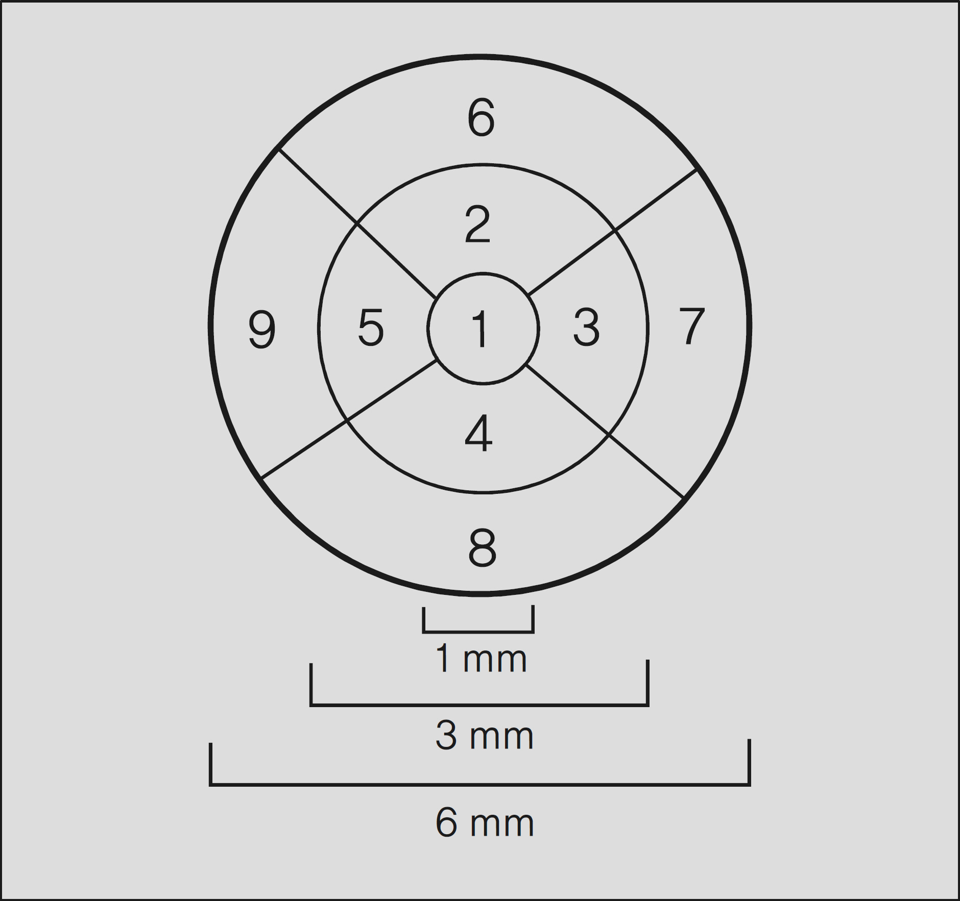
Figure 1. The Age Related Eye Disease Study (AREDS) grid used for reporting retinal thickness and total macular volume in Cirrus HD-OCT system.
Reference
-
References
1. Menke MN, Dabov S, Knecht P, Sturm V. Reproducibility of retinal thickness measurements in healthy subjects using spectralis optical coherence tomography. Am J Ophthalmol. 2009; 147:467–72.
Article2. Nussenblatt RB, Kaufman SC, Palestine AG, et al. Macular thickening and visual acuity. Measurements in patients with cystoid macular edema. Ophthalmology. 1987; 94:1134–9.3. Legarreta JE, Gregori G, Punjabi OS, et al. Macular thickness measurements in normal eyes using spectral domain optical coherence tomography. Ophthalmic Surg Lasers Imaging. 2008; 39:43–9.
Article4. Hogan MJ, Alvarado JA, Weddell JE. Histology of the human eye. Philadelphia: WB Saunders Company;1971. 492.5. Yi K, Chen TC, de Boer JF. Spectral domain optical coherence tomography. Technics in Ophthalmology. 2006; 4:170–4.
Article6. Chen TC, Cense B, Pierce MC, et al. Spectral domain optical coherence tomography: ultra-high speed, ultra-high resolution ophthalmic imaging. Arch Ophthalmol. 2005; 123:1715–20.7. Nassif N, Cense B, Park BH, et al. In vivo human retinal imaging by ultrahigh-speed spectral domain optical coherence tomography. Opt Lett. 2004; 29:480–2.8. Kiernan DF, Hariprasad SM, Chin EK, et al. Prospective comparison of cirrus and stratus optical coherence tomography for quantifying retinal thickness. Am J Ophthalmol. 2009; 147:267–75.
Article9. Age-Related Eye Disease Study Group. The age-related eye disease study severity scale for age-related macular degenereation: AREDS report No. 17. Arch Ophthalmol. 2005; 123:1484–98.10. Zeimer RC, Mori MT, Khoobehi B. Feasibility test of a new method to measure retinal thickness noninvasively. Invest Ophthalmol Vis Sci. 1989; 30:2099–105.11. Gieser JP, Rusin MM, Mori M, et al. Clinical assessment of the macula by retinal topography and thickness mapping. Am J Ophthalmol. 1997; 124:648–60.
Article12. Asrani S, Zou S, d'Anna S, et al. Noninvasive mapping of the normal retinal thickness at the posterior pole. Ophthalmology. 1999; 106:269–73.13. Kohner EM, Dollery CT. Fluorescein angiography of the fundus in diabetic retinopathy. Br Med Bull. 1970; 26:166–70.
Article14. Weinberger D, Axer-Siegel R, Landau D, Yassur Y. Retinal thickness variation in the diabetic patient measured by the retinal thickness analyser. Br J Ophthalmol. 1998; 82:1003–6.
Article15. Fusimoto JG, Brezinski ME, Tearney GJ, et al. Optical biopsy and imaging using optical coherence tomography. Nat Med. 1995; 1:970–2.
Article16. Fusimoto JG, Pitris C, Boppart SA, et al. Optical coherence tomography: an emerging technology for biomedical imaging and optical biopsy. Neoplasia. 2000; 2:9–25.
Article17. Han IC, Jaffe GJ. Comparison of spectral and time domain optical coherence tomography for retinal thickness measurements in healthy and diseased eyes. Am J Ophthalmol. 2009; 147:847–58.18. Kang JH, Kim SA, Song WG, et al. Macular thickness changes with age in normal subjects measured by optical cohrence tomography. J Korean Ophthalmol Soc. 2004; 45:592–8.19. Kanai K, Abe T, Murayama K, et al. Retinal thickness and changes with age. Nippon Ganka Gakkai Zasshi. 2002; 106:162–5.
Article20. Massin P, Duguid G, Erginary A, et al. Optical coherence tomography for evaluating diabetic macular edema before and after vitrectomy. Am J Ophthalmol. 2003; 135:169–77.
Article21. Massin P, Vicaut E, Haouchine B, et al. Reproducibility of retinal mapping using optical coherence tomography. Arch Ophthalmol. 2001; 119:1135–42.
Article22. Hee MR, Puliafito CA, Duker JS, et al. Topography of diabetic macular edema with optical coherence tomography. Ophthalmology. 1998; 105:360–70.
Article23. Jung HJ, Hyun JH, Kim YI, Yun IH. Normal Macular thickness measured macular mapping of OCT3. J Korean Ophthalmol Soc. 2004; 45:962–8.24. Sánchez-Tocino H, Alvarez-Vidal A, Maldonado MJ, et al. Retinal thickness study with optical coherence tomography in patients with diabetes. Invest Ophthalmol Vis Sci. 2002; 43:1588–94.25. Schaudig UH, Glaefke C, Scholz F, Richard G. Optical coherence tomography for retinal thickness measurement in diabetic patients without clinically significant macular edema. Ophthalmic Surg Lasers. 2000; 31:182–6.
Article26. Lee DY, Yu SY, Kwak HW. Quantitative analysis of macular thickness with OCT map. J Korean Ophthalmol Soc. 2004; 45:1496–502.27. Panda-Jonas S, Jonas JB, Jakobczyk-Zmija M. Retinal photo-receptor density decreases with age. Ophthalmology. 1995; 102:1853–9.
Article28. Panda-Jonas S, Jonas JB, Jakobczyk-Zmija M. Retinal pigment epithelial cell count, distribution, and correlations in normal human eyes. Am J Ophthalmol. 1996; 121:181–9.
Article29. Shin JH, Lee HJ, Jin KH. The relationship between axial length, refractive error and foveal thickness measured by OCT in Koreans. J Korean Ophthalmol Soc. 2005; 46:701–6.30. Forooghian F, Cukras C, Meyerle CB, et al. Evaluation of time domain and spectral domain optical coherence tomography in the measurement of diabetic macular edema. Invest Ophthalmol Vis Sci. 2008; 49:4290–6.
Article31. Wakitani Y, Sasoh M, Sugimoto M, et al. Macular thickness measurements in healthy subjects with different axial lengths using optical coherence tomography. Retina. 2003; 23:177–82.
Article
- Full Text Links
-
- Actions
-
Cited
- CITED
-
- Close
- Share
-
- Similar articles
-
- Macular Thickness Changes with Age and Gender in Emmetropia Using Spectral Domain Optical Coherence Tomography
- Repeatability of Spectral Domain OCT (3D-OCT 1000) in Normal Subjects and Various Macular Diseases
- Choroidal Thickness at the Outside of Fovea in Diabetic Retinopathy Using Spectral-Domain Optical Coherence Tomography
- Thickness of the Macula, Retinal Nerve Fiber Layer, and Ganglion Cell-inner Plexiform Layer in the Macular Hole: The Repeatability Study of Spectral-domain Optical Coherence Tomography
- Comparison of Choroidal Thickness in Patients with Diabetes by Spectral-domain Optical Coherence Tomography
