Bronchoscopic Findings of Pulmonary Paragonimiasis
- Affiliations
-
- 1Division of Pulmonary and Critical Care Medicine, Department of Medicine, Sungkyunkwan University School of Medicine, Seoul, Korea. hjk3425@skku.edu
- 2Department of Pathology, Samsung Medical Center, Sungkyunkwan University School of Medicine, Seoul, Korea.
Abstract
- BACKGROUND
Pulmonary paragonimiasis is a subacute to chronic inflammatory disease of the lung caused by lung flukes that result in prolonged inflammation and mechanical injury to the bronchi. However, there are few reports on the bronchoscopic findings of pulmonary paragonimiasis. This report describes the bronchoscopic findings of pulmonary paragonimiasis. METHODS: The bronchosocpic findings of 30 patients (20 males, median age 50 years) with pulmonary paragonimiasis between May 1995 and December 2007 were reviewed retrospectively. RESULTS: The diagnoses were based on a positive serologic test results for Paragonimus-specific antibodies in 13 patients (43%), or the detection of Paragonimus eggs in the sputum, bronchial washing fluid, or lung biopsy specimens in 17 patients (57%). The bronchoscopic examinations revealed endobronchial lesions in 17 patients (57%), which were located within the segmental bronchi in 10 patients (59%), lobar bronchi in 6 patients (35%) and main bronchi in 1 patient (6%). The bronchoscopic characteristics of endobronchial lesions were edematous swelling of the mucosa (16/17, 94%) and mucosal nodularity (4/17, 24%), accompanied by bronchial stenosis in 16 patients (94%). Paragonimus eggs were detected in the bronchial washing fluid of 9 out of the 17 patients with endobronchial lesions. The bronchial mucosal biopsy specimens showed evidence of chronic inflammation with eosinophilic infiltration in 6 out of 11 patients (55%). However, no adult fluke or ova were found in the bronchial tissue. CONCLUSION: Bronchial stenosis with mucosal changes including edematous swelling and mucosal nodularity is the most common bronchoscopic finding of pulmonary paragonimiasis.
Keyword
MeSH Terms
Figure
-
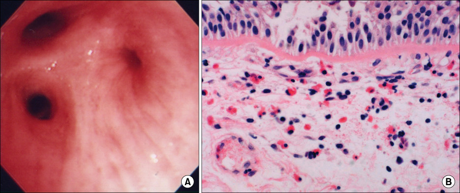
Figure 1 Bronchoscopic appearance and histologic findings of bronchial mucosal biopsy specimens in a 44-year-old male with pulmonary paragonimiasis. (A) Fiberoptic bronchoscopy revealed bronchial stenosis that was associated with mucosal edema of the posterior segmental bronchus of the right upper lobe. (B) Photomicrograph of bronchial tissue obtained from the posterior segmental bronchus of the right upper lobe showing thickening of the basement membrane, chronic inflammation with eosinophilic infiltration, and multinucleated giant cell formation (H&E stain, ×400).

Figure 2 Bronchoscopic appearance and histologic findings of bronchial mucosal biopsy specimens in a 55-year-old female with pulmonary paragonimiasis. (A) Fiberoptic bronchoscopy revealed mucoal nodularity on the right upper lobe. (B) Photomicrograph of bronchial tissue obtained from the right upper lobar bronchus showing thickening of the basement membrane, chronic inflammation with eosinophilic infiltration, and the presence of several multinucleated giant cells (H&E stain, ×200).
Reference
-
1. Im JG, Kong Y, Shin YM, Yang SO, Song JG, Han MC, et al. Pulmonary paragonimiasis: clinical and experimental studies. Radiographics. 1993. 13:575–586.2. Kagawa FT. Pulmonary paragonimiasis. Semin Respir Infect. 1997. 12:149–158.3. Jeon K, Koh WJ, Kim H, Kwon OJ, Kim TS, Lee KS, et al. Clinical features of recently diagnosed pulmonary paragonimiasis in Korea. Chest. 2005. 128:1423–1430.4. Mukae H, Taniguchi H, Matsumoto N, Iiboshi H, Ashitani J, Matsukura S, et al. Clinicoradiologic features of pleuropulmonary Paragonimus westermani on Kyusyu Island, Japan. Chest. 2001. 120:514–520.5. Choi DW. Paragonimus and paragonimiasis in Korea. Korean J Parasitol. 1990. 28:Suppl. 79–102.6. Cho SY, Kong Y, Kang SY. Epidemiology of paragonimiasis in Korea. Southeast Asian J Trop Med Public Health. 1997. 28:Suppl 1. 32–36.7. Yokogawa M. Paragonimus and paragonimiasis. Adv Parasitol. 1969. 7:375–387.8. Rangdaeng S, Alpert LC, Khiyami A, Cottingham K, Ramzy I. Pulmonary paragonimiasis: report of a case with diagnosis by fine needle aspiration cytology. Acta Cytol. 1992. 36:31–36.9. Im JG, Whang HY, Kim WS, Han MC, Shim YS, Cho SY. Pleuropulmonary paragonimiasis: radiologic findings in 71 patients. AJR Am J Roentgenol. 1992. 159:39–43.10. Diaconita G, Goldis G. Pathomorphology and pathogenesis of pulmonary paragonimiasis. Acta Morphol Acad Sci Hung. 1964. 12:315–331.11. Yoshino I, Nawa Y, Yano T, Ichinose Y. Paragonimiasis westermani presenting as an asymptomatic nodular lesion in the lung: report of a case. Surg Today. 1998. 28:108–110.12. Tomita M, Matsuzaki Y, Nawa Y, Onitsuka T. Pulmonary paragonimiasis referred to the department of surgery. Ann Thorac Cardiovasc Surg. 2000. 6:295–298.
