Restor Dent Endod.
2013 Nov;38(4):204-209.
A comparative evaluation of cytotoxicity of root canal sealers: an in vitro study
- Affiliations
-
- 1Department of Conservative Dentistry & Endodontics VSPM's Dental College & Research Center, Nagpur, India. badole_g15@yahoo.co.in
- 2Department of Conservative Dentistry & Endodontics Government Dental College & Hospital, Nagpur, India.
- 3Department of Conservative Dentistry & Endodontics, Peoples Dental College, Bhopal, India.
- 4Department of Pedodontics, Sharad Pawar Dental College, Sawangi, Wardha, India.
Abstract
OBJECTIVES
The objective of this in vitro study was to evaluate and compare the cytotoxicity of four different root canal sealers i.e. Apexit Plus (Ivoclar Vivadent), Endomethasone N (Septodont), AH-26 (Dentsply) and Pulpdent Root Canal Sealer (Pulpdent), on a mouse fibroblast cell line (L929).
MATERIALS AND METHODS
Thirty two discs for each sealer (5 mm in diameter and 2 mm in height) were fabricated in Teflon mould. The sealer extraction was made in cell culture medium (Dulbecco's Modified Eagle's Medium, DMEM) using the ratio 1.25 cm2/mL between the surface of the sealer samples and the volume of medium in a shaker incubator. Extraction of each sealer was obtained at 24 hr, 7th day, 14th day, and one month of interval. These extracts were incubated with L929 cell line and 3-(4,5-dimethylthiazol-2yl)-2,5-diphenyltetrazolium bromide (MTT) assay was done. Two-way ANOVA for interaction effects between sealer and time and Post-hoc multiple comparison using Tukey's test across all the 16 different groups were used for statistical analysis.
RESULTS
Apexit Plus root canal sealer was significantly less toxic than other sealers (p < 0.05) and showed higher cellular growth than control. Endomethasone N showed mild cytotoxicity. AH-26 showed severe toxicity which became mild after one month while Pulpdent Root Canal Sealer showed severe to moderate toxicity.
CONCLUSIONS
Apexit Plus was relatively biocompatible sealer as compared to other three sealers which were cytotoxic at their initial stages, however, they became biocompatible with time.
Keyword
MeSH Terms
-
Animals
Bismuth
Calcium Hydroxide
Cell Culture Techniques
Cell Line
Dental Pulp Cavity*
Dexamethasone
Drug Combinations
Epoxy Resins
Fibroblasts
Formaldehyde
Hydrocortisone
Incubators
Mice
Polytetrafluoroethylene
Root Canal Filling Materials
Silver
Thymol
Titanium
Bismuth
Calcium Hydroxide
Dexamethasone
Drug Combinations
Epoxy Resins
Formaldehyde
Hydrocortisone
Polytetrafluoroethylene
Root Canal Filling Materials
Silver
Thymol
Titanium
Figure
-
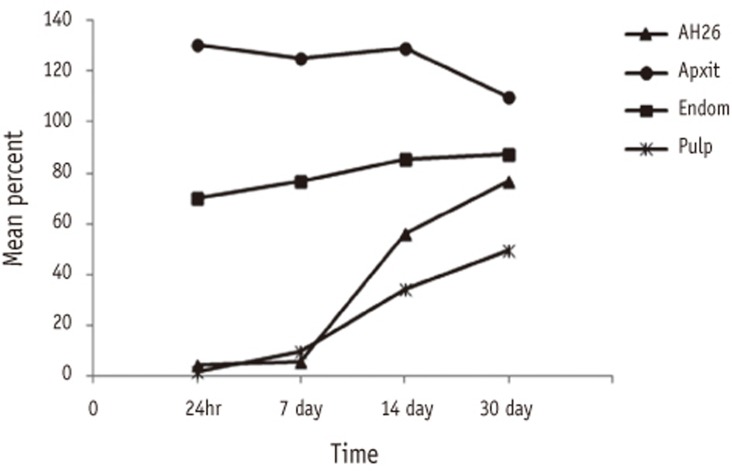
Figure 1 Percentages of cell viability in four experimental groups demonstrated significant interaction effects between sealer and time.
Reference
-
1. Ingle JI, Backland KL. Chapter 30, Obturation of the radicular space. Endodontics. 6th ed. Hamilton: BC Decker Inc.;2008. p. 1053–1087.2. Grossman LI, Oliet S, Del Rio CE. Chapter 9, Obturation of radicular space. Endodontic practice. 12th ed. New Delhi: Walter Kluwer Pvt. Ltd.;2010. p. 278–309.3. De Deus QD. Frequency, location, and direction of lateral, secondary, and accessory canals. J Endod. 1975; 1:361–366. PMID: 10697487.4. Dongari A, Lambrianidis T. Periodontally derived pulpal lesions. Endod Dent Traumatol. 1988; 4:49–54. PMID: 3251755.
Article5. Dahl JE, Frangou-Polyzois MJ, Polyzois GL. In vitro biocompatibility of denture relining materials. Gerodontology. 2006; 23:17–22. PMID: 16433637.6. Miletić I, Devcić N, Anić I, Borcić J, Karlović Z, Osmak M. The cytotoxicity of Roeko Seal and AH plus compared during different setting periods. J Endod. 2005; 31:307–309. PMID: 15793391.7. Al-Nazhan S, Spangberg L. Morphological cell changes due to chemical toxicity of a dental material: an electron microscopic study on human periodontal ligament fibroblasts and L929 cells. J Endod. 1990; 16:129–134. PMID: 2388028.
Article8. ISO-Standards ISO 10993. Biological compatibility of medical devices. Test for cytotoxicity: in vitro methods. part 5. International Organization for Standardization;2009. cited 2013 July 9. https://law.resource.org/pub/ie/ibr/is.en.iso.10993.5.2009.html.9. Leonardo RT, Consolaro A, Carlos IZ, Leonardo MR. Evaluation of cell culture cytotoxicity of five root canal sealers. J Endod. 2000; 26:328–330. PMID: 11199748.
Article10. Huang FM, Hsieh YS, Tai KW, Chou MY, Chang YC. Induction of c-fos and c-jun protooncogenes expression by formaldehyde-releasing and epoxy resin-based root-canal sealers in human osteoblastic cells. J Biomed Mater Res. 2002; 59:460–465. PMID: 11774303.
Article11. Mosmann T. Rapid colorimetric assay for cellular growth and survival: application to proliferation and cytotoxicity assays. J Immunol Methods. 1983; 65:55–63. PMID: 6606682.
Article12. Oztan MD, Yilmaz S, Kalayci A, Zaimoğlu A. A comparison of the in vitro cytotoxicity of two root canal sealers. J Oral Rehabil. 2003; 30:426–429. PMID: 12631168.13. Koulaouzidou EA, Papazisis KT, Beltes P, Geromichalos GD, Kortsaris AH. Cytotoxicity of three resin-based root canal sealers: an in vitro evaluation. Endod Dent Traumatol. 1998; 14:182–185. PMID: 9796482.14. Pinna L, Brackett MG, Lockwood PE, Huffman BP, Mai S, Cotti E, Dettori C, Pashley DH, Tay FR. In vitro cytotoxicity evaluation of a self-adhesive, methacrylate resin-based root canal sealer. J Endod. 2008; 34:1085–1088. PMID: 18718370.15. Osorio RM, Hefti A, Vertucci FJ, Shawley AL. Cytotoxicity of endodontic materials. J Endod. 1998; 24:91–96. PMID: 9641138.
Article16. Azar NG, Heidari M, Bahrami ZS, Shokri F. In vitro cytotoxicity of a new epoxy resin root canal sealer. J Endod. 2000; 26:462–465. PMID: 11199780.17. Miletić I, Anić I, Karlović Z, Marsan T, Pezelj-Ribarić S, Osmak M. Cytotoxic effect of four root filling materials. Endod Dent Traumatol. 2000; 16:287–290. PMID: 11202896.
Article18. Geurtsen W, Leinenbach F, Krage T, Leyhausen G. Cytotoxicity of four root canal sealers in permanent 3T3 cells and primary human periodontal ligament fibroblast cultures. Oral Surg Oral Med Oral Pathol Oral Radiol Endod. 1998; 85:592–597. PMID: 9619680.
Article19. Nakamura H, Sakakibara F, Matsumoto Y, Hirano S, Hayakawa H, Sakai K, Yip M. Study on cytotoxicity of root canal filling materials. J Endod. 1986; 12:156–160. PMID: 3461103.20. Guigand M, Pellen-Mussi P, Le Goff A, Vulcain JM, Bonnaure-Mallet M. Evaluation of the cytotocompatibility of three endodontic materials. J Endod. 1999; 25:419–423. PMID: 10530242.21. Gerosa R, Menegazzi G, Borin M, Cavalleri G. Cytotoxicity evaluation of six root canal sealers. J Endod. 1995; 21:446–448. PMID: 8537785.
Article22. Schwarze T, Fiedler I, Leyhausen G, Geurtsen W. The cellular compatibility of five endodontic sealers during the setting period. J Endod. 2002; 28:784–786. PMID: 12470025.
Article23. Torneck CD, Moe H, Howley TP. The effect of calcium hydroxide on porcine pulp fibroblasts in vitro. J Endod. 1983; 9:131–136. PMID: 6574198.24. Swierenga SH, MacManus JP, Whitfield JF. Regulation by calcium of the proliferation of heart cells from young adult rats. In Vitro. 1976; 12:31–36. PMID: 942664.
Article25. Whitfield JF, MacManus JP, Rixon RH, Boynton AL, Yondale T, Swierenga S. The positive control of cell proliferation by the interplay on calcium ions and cyclic nucleotides. A review. In Vitro. 1976; 12:1–18. PMID: 172436.26. Kim CK, Ryu HW, Chang HS, Lee BD, Min KS, Hong CU. Evaluation of the radiopacity and cytotoxicity of resinous root canal sealers. J Korean Acad Conserv Dent. 2007; 32:419–425.
Article27. Schwarze T, Leyhausen G, Geursten W. Long-term cytocompatibility of various endodontic sealers using a new root canal model. J Endod. 2002; 28:749–753. PMID: 12470017.
Article28. Kim HJ, Baek SH, Bae KS. Cytotoxicity and genotoxicity of newly developed calcium phosphate-based root canal sealers. J Korean Acad Conserv Dent. 2006; 31:36–49.
Article29. Park SY, Lee WC, Lim SS. Cytotoxicity and antibacterial property of new resin-based sealers. J Korean Acad Conserv Dent. 2003; 28:162–168.30. Cohen BI, Pagnillo MK, Musikant BL, Deutsch AS. An in vitro study of the cytotoxicity of two root canal sealers. J Endod. 2000; 26:228–229. PMID: 11199724.31. Vajrabhaya L, Sithisarn P. Multilayer and monolayer cell cultures in a cytotoxicity assay of root canal sealers. Int Endod J. 1997; 30:141–144. PMID: 10332248.
Article32. Spångberg LS, Barbosa SV, Lavigne GD. AH-26 releases formaldehyde. J Endod. 1993; 19:596–598. PMID: 8151253.
Article33. Kaplan AE, Goldberg F, Artaza LP, de Silvio A, Macchi RL. Disintegration of endodontic cements in water. J Endod. 1997; 23:439–441. PMID: 9587297.
Article34. Araki K, Suda H, Barbosa SV, Spångberg LS. Reduced cytotoxicity of a root canal sealer through eugenol substitution. J Endod. 1993; 19:554–557. PMID: 8151243.
Article35. Key JE, Rahemtulla FG, Eleazer PD. Cytotoxicity of a new root canal filling material on human gingival fibroblasts. J Endod. 2006; 32:756–758. PMID: 16861076.
Article36. Watts A, Paterson RC. Cellular responses in the dental pulp: a review. Int Endod J. 1981; 14:10–19. PMID: 7024136.
Article37. Wataha JC, Hanks CT, Strawn SE, Fat JC. Cytotoxicity of components of resins and other dental restorative materials. J Oral Rehabil. 1994; 21:453–462. PMID: 7965356.
Article38. Wiegand C, Hipler UC. Methods for the measurement of cell and tissue compatibility including tissue regeneration processes. GMS Krankenhhyg Interdiszip. 2008; 3:Doc12. PMID: 20204114.
- Full Text Links
-
- Actions
-
Cited
- CITED
-
- Close
- Share
-
- Similar articles
-
- Cytotoxicity of resin-based root canal sealer, adseal
- Evaluation of the radiopacity and cytotoxicity of resinous root canal sealers
- Calcium silicate-based root canal sealers: a literature review
- Cytotoxicity and genotoxicity of newly developed calcium phosphate-based root canal sealers
- The status of clinical trials regarding root canal sealers
