Two Cases of Post-Radiation Sarcoma after Breast Cancer Treatment
- Affiliations
-
- 1Department of Radiation Oncology, Samsung Medical Center, Sungkyunkwan University School of Medicine, Seoul, Korea. sj5201.huh@samsung.com
- 2Department of Surgery, Samsung Medical Center, Sungkyunkwan University School of Medicine, Seoul, Korea.
- KMID: 2286465
- DOI: http://doi.org/10.4048/jbc.2012.15.3.364
Abstract
- We describe two cases of post-radiation sarcoma after breast cancer treatment. The first patient was a 61-year-old woman who underwent partial mastectomy of the right breast and adjuvant whole breast irradiation 7 years previously. Subsequently, a rapidly growing mass from the anterior arc of the right fifth rib was incidentally detected on an abdomino-pelvic computed tomography scan. The second patient was a 70-year-old woman who received neoadjuvant chemotherapy and a partial mastectomy of the left breast 9 years ago. Adjuvant irradiation was delivered to the whole breast and supraclavicular region. Subsequently, an approximate 8 cm mass developed in the left axillary area. Both patients received wide excision of the tumor with negative resection margins. The pathological diagnoses were osteosarcoma and undifferentiated pleomorphic sarcoma, respectively. Although post-radiation sarcomas are rare complications with a poor prognosis, enhanced awareness and early detection by clinicians are essential to improve outcomes via curative surgical resection.
MeSH Terms
Figure
-
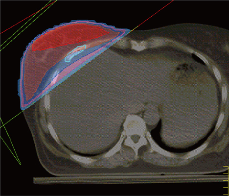
Figure 1 The previously used irradiation fields in case 1. Total doses delivered were 50, 45, and 40 Gy to the areas filled with red, blue, and pink, respectively. The fifth rib, where post-radiation sarcoma developed, is delineated in sky blue.
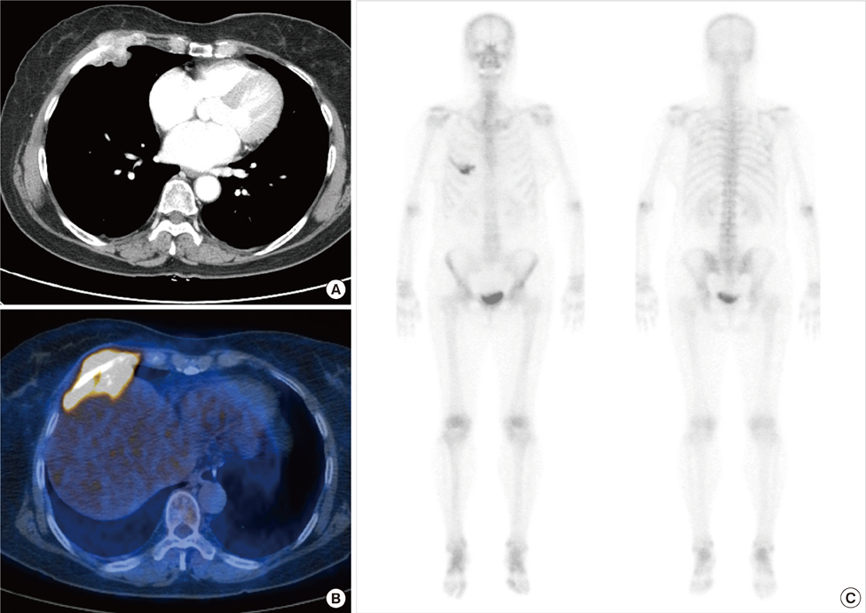
Figure 2 Imaging studies of case 1. An enhancing mass at the anterior arc of the right fifth rib was discovered on a computed tomography scan (A). The mass was hypermetabolic on a positron emission tomography scan (B) and increased radio-uptake was shown on a whole body bone scan (C).
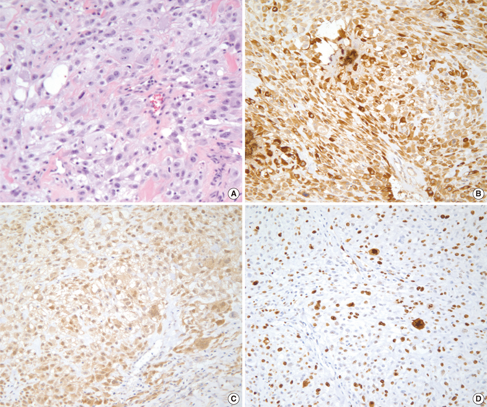
Figure 3 Histological examination of case 1. (A) Atypical tumor cells with osteoid production are shown on the specimen (H&E, ×200). The tumor revealed positive immunoreactivity for osteocalcin (B), cyclin-dependent kinase 4 (C), and Ki-67 (D, 60%) (immunohistochemical staining, ×200).
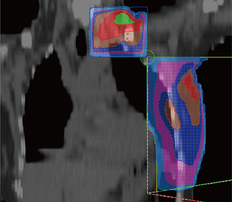
Figure 4 Coronal reconstruction of the previously used irradiation fields of case 2. The dose distributions of the filled areas are the same as those in Figure 1.
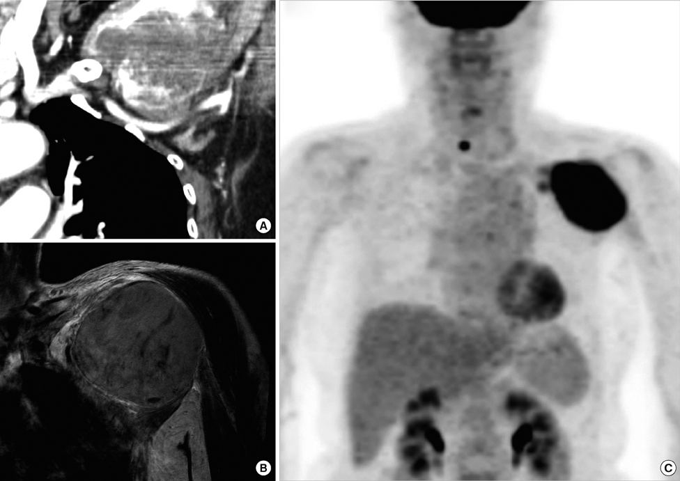
Figure 5 Imaging studies of case 2. An approximate 8-cm mass with peripheral enhancement developed in the left axillary area was observed on computed tomography (A) and magnetic resonance imaging scans (B). The mass was also hypermetabolic on a positron emission tomography scan (C).
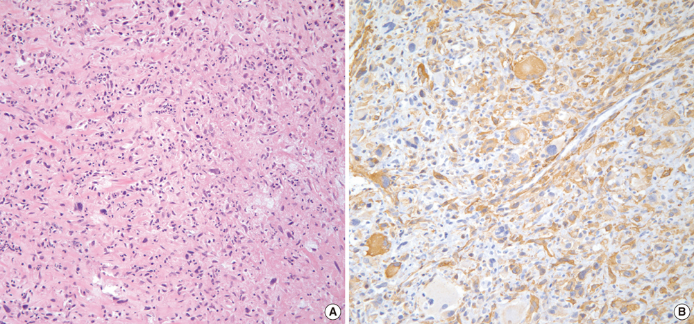
Figure 6 Histological examination of case 2. (A) Many atypical tumor cells were seen in the specimen (H&E, ×200). (B) The tumor had positive immunoreactivity for smooth muscle actin (immunohistochemical staining, ×200).
Reference
-
1. Cahan WG, Woodard HQ, Higinbotham NL, Stewart FW, Coley BL. Sarcoma arising in irradiated bone: report of 11 cases. Cancer. 1948. 1:3–29.2. Murray EM, Werner D, Greeff EA, Taylor DA. Postradiation sarcomas: 20 cases and a literature review. Int J Radiat Oncol Biol Phys. 1999. 45:951–961.
Article3. Sheppard DG, Libshitz HI. Post-radiation sarcomas: a review of the clinical and imaging features in 63 cases. Clin Radiol. 2001. 56:22–29.
Article4. Veronesi U, Cascinelli N, Mariani L, Greco M, Saccozzi R, Luini A, et al. Twenty-year follow-up of a randomized study comparing breast-conserving surgery with radical mastectomy for early breast cancer. N Engl J Med. 2002. 347:1227–1232.
Article5. Yap J, Chuba PJ, Thomas R, Aref A, Lucas D, Severson RK, et al. Sarcoma as a second malignancy after treatment for breast cancer. Int J Radiat Oncol Biol Phys. 2002. 52:1231–1237.
Article6. Kirova YM, Vilcoq JR, Asselain B, Sastre-Garau X, Fourquet A. Radiation-induced sarcomas after radiotherapy for breast carcinoma: a large-scale single-institution review. Cancer. 2005. 104:856–863.
Article7. Erel E, Vlachou E, Athanasiadou M, Hassan S, Chandrasekar CR, Peart F. Management of radiation-induced sarcomas in a tertiary referral centre: a review of 25 cases. Breast. 2010. 19:424–427.
Article8. Sheth GR, Cranmer LD, Smith BD, Grasso-Lebeau L, Lang JE. Radiation-induced sarcoma of the breast: a systematic review. Oncologist. 2012. 17:405–418.
Article9. Acharya S, Sarafoglou K, LaQuaglia M, Lindsley S, Gerald W, Wollner N, et al. Thyroid neoplasms after therapeutic radiation for malignancies during childhood or adolescence. Cancer. 2003. 97:2397–2403.
Article10. Huang J, Walker R, Groome PG, Shelley W, Mackillop WJ. Risk of thyroid carcinoma in a female population after radiotherapy for breast carcinoma. Cancer. 2001. 92:1411–1418.
Article11. Kirova YM, Gambotti L, De Rycke Y, Vilcoq JR, Asselain B, Fourquet A. Risk of second malignancies after adjuvant radiotherapy for breast cancer: a large-scale, single-institution review. Int J Radiat Oncol Biol Phys. 2007. 68:359–363.
Article12. Rubino C, Shamsaldin A, Lê MG, Labbé M, Guinebretière JM, Chavaudra J, et al. Radiation dose and risk of soft tissue and bone sarcoma after breast cancer treatment. Breast Cancer Res Treat. 2005. 89:277–288.
Article13. Krueger EA, Fraass BA, McShan DL, Marsh R, Pierce LJ. Potential gains for irradiation of chest wall and regional nodes with intensity modulated radiotherapy. Int J Radiat Oncol Biol Phys. 2003. 56:1023–1037.
Article14. Tabone MD, Terrier P, Pacquement H, Brunat-Mentigny M, Schmitt C, Babin-Boilletot A, et al. Outcome of radiation-related osteosarcoma after treatment of childhood and adolescent cancer: a study of 23 cases. J Clin Oncol. 1999. 17:2789–2795.
Article15. Jung YS, Na KY, Kim KS, Ahn SH, Lee SJ, Park HK, et al. Nation-wide Korean breast cancer data from 2008 using the breast cancer registration program. J Breast Cancer. 2011. 14:229–236.
Article
- Full Text Links
-
- Actions
-
Cited
- CITED
-
- Close
- Share
-
- Similar articles
-
- Post Radiation Myxofibrosarcoma in Breast: A Case Report
- Breast Cancer after Radiation Therapy in a Patient with Li-Fraumeni Syndrome: A Case Report
- Sinonasal Undifferentiated Pleomorphic Sarcoma in Five Patient Cases
- Ultrasonographic Findings of Post-Operative Changes after Breast Cancer Surgery
- A Radiation Induced Low-Grade Myofibroblastic Sarcoma in the Retropectoral Area After Breast Conserving Surgery: A Case Report
