Post Radiation Myxofibrosarcoma in Breast: A Case Report
- Affiliations
-
- 1Department of Radiology, College of Medicine, Yeungnam University, Daegu, Korea. raina_eleven@hanmail.net
- 2Department of Pathology, College of Medicine, Yeungnam University, Daegu, Korea.
- 3Department of General Surgery, College of Medicine, Yeungnam University, Daegu, Korea.
- KMID: 1823924
- DOI: http://doi.org/10.3348/jksr.2015.73.1.53
Abstract
- Post radiation sarcoma is a very rare and invasive malignant tumor that occurs in patients who had radiation therapy 5-10 years ago for primary cancer such as breast cancer, lung cancer or lymphoma. In recent years, the incidence of post radiation sarcoma has increased, as patients survive longer after radiation therapy. We reported a case of surgically confirmed post radiation myxofibrosarcoma from the breast of a 59-year-old woman, with a discussion on the literature.
Figure
-
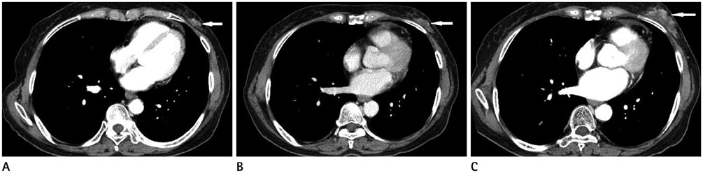
Fig. 1 CT images of post radiation sarcoma. A. On initial CT scan, irregular margined soft tissue lesion (arrow) is seen in left breast outer lower quadrant with similar attenuation of adjacent muscle. B. At 3 months from initial CT, the soft tissue lesion (arrow) is enlarged, as compared to the previous image and shows subtle enhancement of tumor margin. C. At 5 months from initial CT, this lesion (arrow) is further enlarged and shows markedly irregular margins and heterogeneous enhancement.
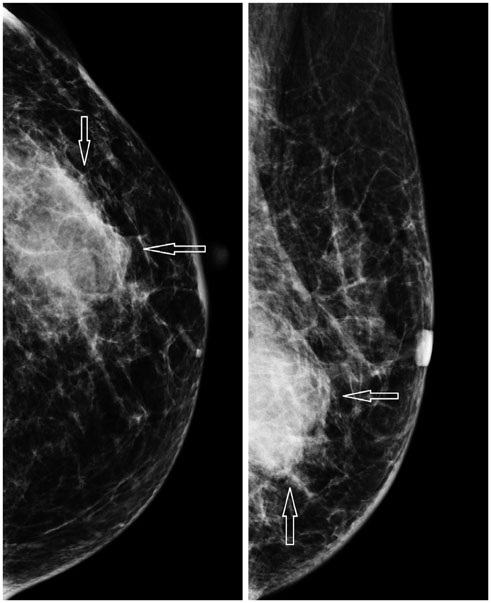
Fig. 2 Mammography images of post radiation sarcoma. At approximately 7 month after the initial CT scan, mammography is performed. Background breast shows heterogeneous density. About 4 cm sized mass is seen in the lower outer quadrant of the left breast in the 5 o'clock position. The mass is hyperdense and shows relatively irregular shape and indistinct margin with partial spiculation. However, calcification or architectural distortion is not seen (arrows).
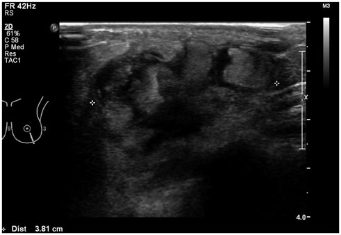
Fig. 3 Ultrasonography image of post radiation sarcoma. One week after the mammography scan, breast ultrasonography is performed. About 3.8 cm size heterogeneous hypoechoic lesion is seen in the left breast outer lower quadrant. Mass shows irregular shape and somewhat parallel orientation. In addition, the margin of the mass is indistinct in principle, but some portion shows a lobulated border.
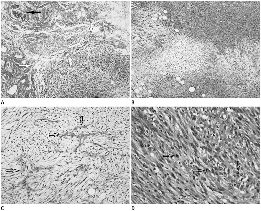
Fig. 4 Histopathologic image of surgically resected specimen. A. Diffuse malignant cells (white arrows) that is infiltrative to adjacent normal tissue is seen in the right lower part and normal breast duct structure (black arrow) is seen in left upper part. Thus, the sarcoma appears to originate from breast tissue, and not another origin (H&E stain, × 100). B. Malignant cells are composed of spindle cells with relatively high cellular density (top and bottom of image) and myxoid portion with relatively low cellular density (middle of image) (H&E stain, × 100). C. In the myxoid portion, multiple curvilinear shaped capillary vasculature (arrows) are seen (H&E stain, × 400). D. In the high cellular density portion, numerous spindle cells show moderate to high grade polymorphism, and some show mitotic events (arrows) in the cell nucleus that are suggestive that the sarcoma is a high grade tumor (H&E stain, × 400). H&E = hematoxylin and eosin
Reference
-
1. Sheth GR, Cranmer LD, Smith BD, Grasso-Lebeau L, Lang JE. Radiation-induced sarcoma of the breast: a systematic review. Oncologist. 2012; 17:405–418.2. Labidi-Galy SI, Tassy L, Blay JY. Radiation induced soft tissue sarcoma. ESUN. 2011; 8:1–12. Available from: http://sarcomahelp.org/radiation-induced-sarcoma.html.3. Penel N, Grosjean J, Robin YM, Vanseymortier L, Clisant S, Adenis A. Frequency of certain established risk factors in soft tissue sarcomas in adults: a prospective descriptive study of 658 cases. Sarcoma. 2008; 2008:459386. [Epub]. DOI: 10.1155/2008/459386.4. Chahin F, Paramesh A, Dwivedi A, Peralta R, O'Malley B, Washington T, et al. Angiosarcoma of the breast following breast preservation therapy and local radiation therapy for breast cancer. Breast J. 2001; 7:120–123.5. Sheppard DG, Libshitz HI. Post-radiation sarcomas: a review of the clinical and imaging features in 63 cases. Clin Radiol. 2001; 56:22–29.6. Cai PQ, Wu YP, Li L, Zhang R, Xie CM, Wu PH, et al. CT and MRI of radiation-induced sarcomas of the head and neck following radiotherapy for nasopharyngeal carcinoma. Clin Radiol. 2013; 68:683–689.7. Gladdy RA, Qin LX, Moraco N, Edgar MA, Antonescu CR, Alektiar KM, et al. Do radiation-associated soft tissue sarcomas have the same prognosis as sporadic soft tissue sarcomas? J Clin Oncol. 2010; 28:2064–2069.8. Bjerkehagen B, Smeland S, Walberg L, Skjeldal S, Hall KS, Nesland JM, et al. Radiation-induced sarcoma: 25-year experience from the Norwegian Radium Hospital. Acta Oncol. 2008; 47:1475–1482.9. Neuhaus SJ, Pinnock N, Giblin V, Fisher C, Thway K, Thomas JM, et al. Treatment and outcome of radiation-induced soft-tissue sarcomas at a specialist institution. Eur J Surg Oncol. 2009; 35:654–659.10. Lagrange JL, Ramaioli A, Chateau MC, Marchal C, Resbeut M, Richaud P, et al. Sarcoma after radiation therapy: retrospective multiinstitutional study of 80 histologically confirmed cases. Radiation Therapist and Pathologist Groups of the Fédération Nationale des Centres de Lutte Contre le Cancer. Radiology. 2000; 216:197–205.
