J Rhinol.
2016 Nov;23(2):115-118. 10.18787/jr.2016.23.2.115.
Sinonasal Undifferentiated Pleomorphic Sarcoma in Five Patient Cases
- Affiliations
-
- 1Department of Otolaryngology, Asan Medical Center, University of Ulsan College of Medicine, Seoul, Korea. jhkim0217@gmail.com
- KMID: 2361325
- DOI: http://doi.org/10.18787/jr.2016.23.2.115
Abstract
- Undifferentiated pleomorphic sarcoma (UPS) is a rare soft tissue sarcoma of the sinonasal area. Here, we present two primary cases of UPS and three post-irradiation sinonasal UPS cases. Imaging findings were misinterpreted by radiologists as representing other malignant tumors or recurrence of the primary tumor. Our cases indicate that post-irradiation UPS can originate within any part of the radiation field. Treatment outcomes of primary sinonasal UPS seem to be favorable if the tumor is treated aggressively, but the outcomes of post-irradiation sinonasal UPS may be poor if appropriate surgical margins cannot be obtained.
MeSH Terms
Figure
-
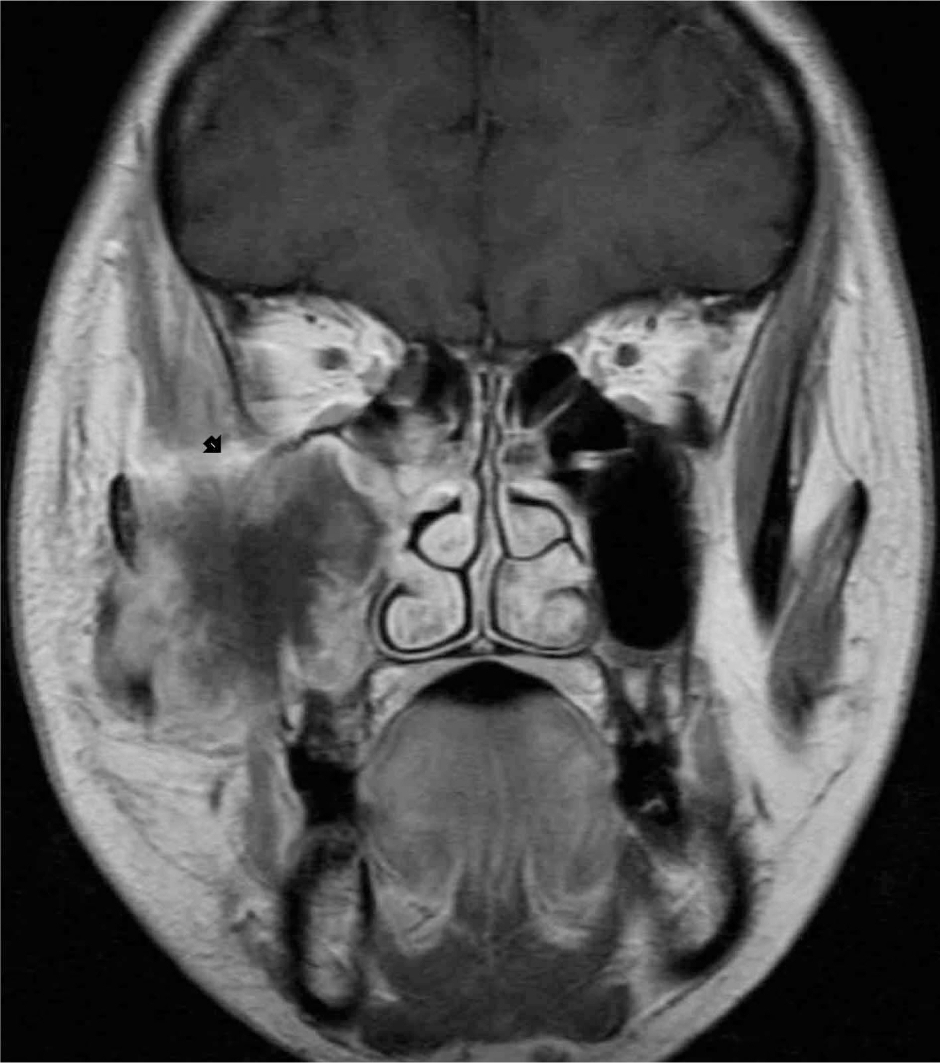
Fig. 1. A bulky soft tissue mass in the right maxillary sinus with destruction of the maxillary walls and extension to the buccal space in gadolinium-enhanced T1-weighted MR image of primary UPS.
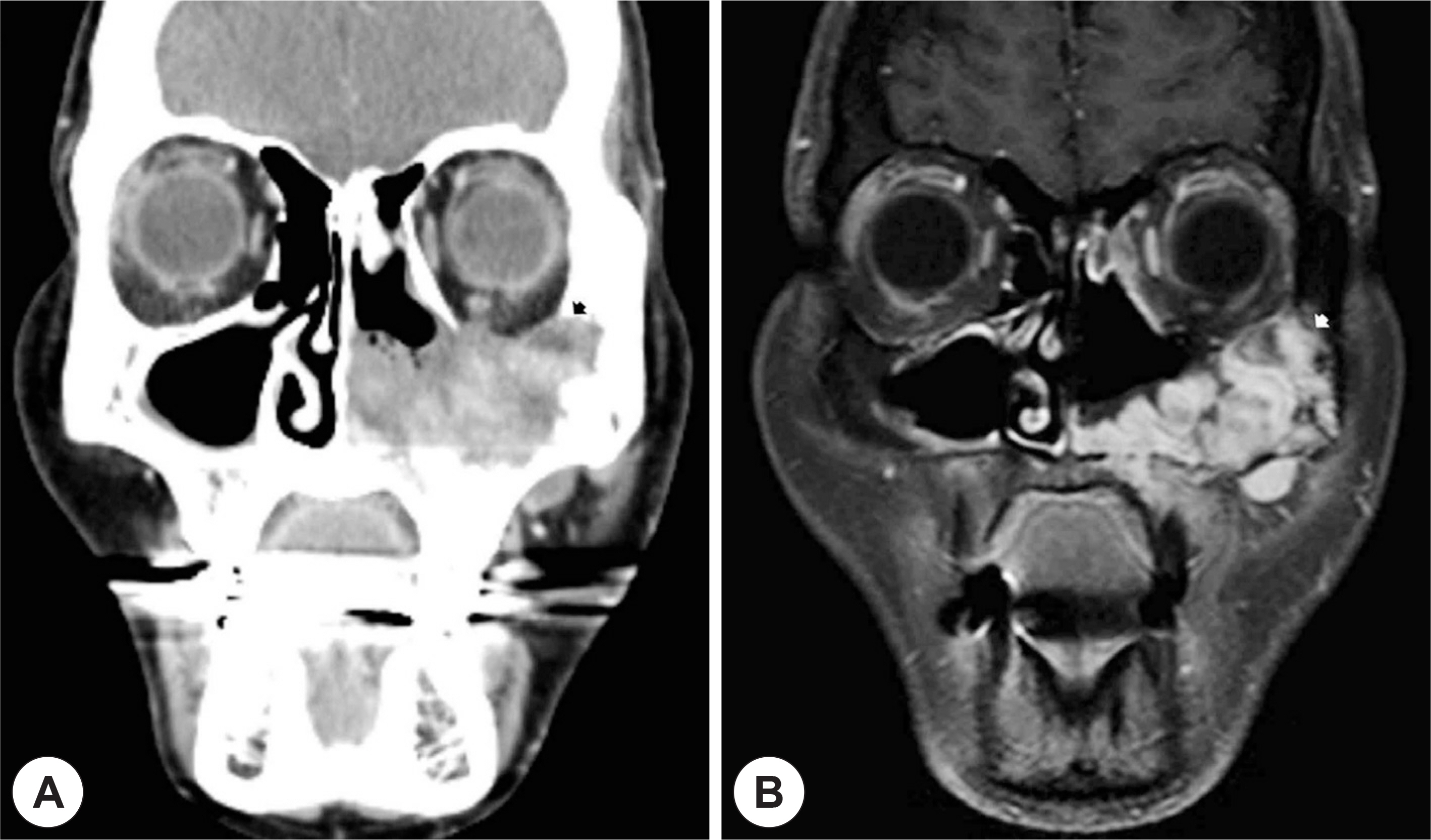
Fig. 2. Heterogeneously enhancing large mass with bony destruction of the maxillary walls and nasal cavity in CT scan (A) and MR image (B) of post-irradiated UPS.
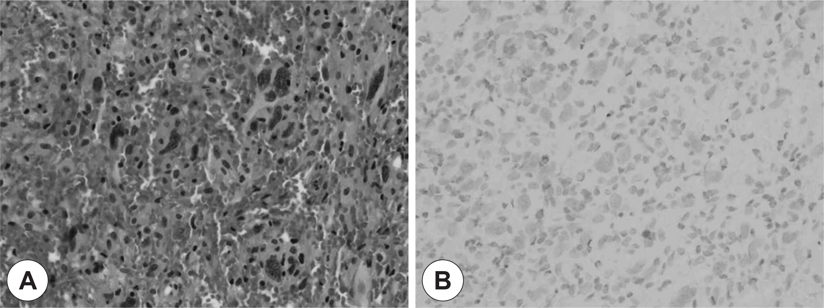
Fig. 3. Histopathology of radiation-induced undifferentiated pleomorphic sarcoma. The tumor consists of mixed spindle cells and roundedcells arranged in a stori-form pattern. A: Hematoxylin-eo-sin (×400). B: Negative staining for cytokeratin by immunohistochemistry (×400).
Reference
-
References
1). Weiss SW, Enzinger FM. Malignant fibrous histiocytoma: an analysis of 200 cases. Cancer. 1978; 41:2250–66.2). Barnes L, Kanbour A. Malignant fibrous histiocytoma of the head and neck. A report of 12 cases. Arch Otolaryngol Head Neck Surg. 1988; 114:1149–56.
Article3). Park SW, Kim HJ, Lee JH, Ko YH. Malignant fibrous histiocytoma of the head and neck: CT and MR imaging findings. AJNR Am J Neuroradiol. 2009; 30:71–6.
Article4). Wang CP, Chang YL, Ting LL, Yang TL, Ko JY, Lou PJ. Malignant fibrous histiocytoma of the sinonasal tract. Head & Neck. 2009; 31:85–93.
Article5). Sheppard DG, Libshitz HI. Postradiation sarcomas: a review of the clinical and imaging features in 63 cases. Clinical Radiology. 2001; 56:22–9.
Article6). Arlen M, Higinbotham NL, Huvos AG, Marcove RC, Miller T, Shah IC. Radiation-induced sarcoma of bone. Cancer. 1971; 28:1087–99.
Article7). Goldblum JR, Folpe AL, Weiss SW. Undifferentiated Pleomorphic Sarcoma. Goldblum JR, editor. editor.Enzinger and Weiss's Soft Tissue Tumors. 6th ed.Philadelphia: Elsevier;2013. p. 421–40.8). Guillou L, Folpe AL. Fibroblastic and Fibrohistiocytic Tumors. Folpe AL, editor. editor.Bone and Soft Tissue Pathology. Philadelphia: Saunders & Elsevier;2010. p. 43–96.
Article9). Agaimy A, Gaumann A, Schroeder J, Dietmaier W, Hartmann A, Hofstaedter F, et al. Primary and metastatic high-grade pleomorphic sarcoma/malignant fibrous histiocytoma of the gastrointestinal tract: an approach to the differential diagnosis in a series of five cases with emphasis on myofibroblastic differentiation. Virchows Arch. 2007; 451:949–57.
Article10). Shahoon H, Esmaeili M, Nematollahi M. Eight-year Follow-up of Malignant Fibrous Histiocytoma (Undifferentiated High-grade Pleomorphic Sarcoma) of the Maxilla: Case Report and Review of the Literature. J Dent Res Dent Clin Dent Prospects. 2009; 3:32–5.11). Vuity D, Bogdan S, Csurgay K, Sapi Z. Malignant fibrous histiocy-toma/undifferentiated high-grade pleomorphic sarcoma of the maxillary sinus: report of a case and review of the literature. Pathol Oncol Res. 2013; 19:605–9.12). Blitzer A, Lawson W, Zak FG, Biller HF, Soon ML. Clinical-pathological determinants in prognosis of fibrous histiocytomas of head and neck. Laryngoscope. 1981; 91:2053–70.
Article13). Weber RS, Benjamin RS, Peters LJ, Ro JY, Achon O, Goepfert H. Soft tissue sarcomas of the head and neck in adolescents and adults. Am J Surg. 1986; 152:386–92.
Article14). Rodrigo JP1. Fernández JA, Suárez C, Gómez J, Llorente JL, Herrero A. Malignant fibrous histiocytoma of the nasal cavity and paranasal sinuses. Am J Rhinol. 2000; 14:427–31.15). Park SW, Kim HJ, Lee JH, Ko YH. Malignant fibrous histiocytoma of the head and neck: CT and MR imaging findings. AJNR Am J Neuroradiol. 2009; 30:71–6.
Article16). Bentz BG, Singh B, Woodruff J, Brennan M, Shah JP, Kraus D. Head and neck soft tissue sarcomas: a multivariate analysis of outcomes. Ann Surg Oncol. 2004; 11:619–28.
Article17). Jang TY, Kim CH, Kim YM, Chu YC. Two cases of malignant fibrous histiocytoma in the nasal cavity and paranasal sinus. J Rhinol. 1998; 5:72–5.18). Seo BS, Choi SJ, Jang YJ, Chung YS, Lee BJ. Recurrence patterns of the maxillary sinus cancer after total maxillectomy. J Rhinol. 2008; 15:39–43.19). Hsu HC, Huang EY, Wang CJ. Treatment results and prognostic factors in patients with malignant fibrous histiocytoma. Acta Oncol. 2004; 43:530–5.
Article20). Le Doussal V, Coindre JM, Leroux A, Hacene K, Terrier P, Bui NB, et al. Prognostic factors for patients with localized primary malignant fibrous histiocytoma: a multicenter study of 216 patients with multivariate analysis. Cancer. 1996; 77:1823–30.
Article
- Full Text Links
-
- Actions
-
Cited
- CITED
-
- Close
- Share
-
- Similar articles
-
- A Case of Pleomorphic Dermal Sarcoma Showing Characteristics of Myxoinflammatory Fibroblastic Sarcoma
- Cutaneous Metastatic Undifferentiated Pleomorphic Sarcoma from a Mediastinal Sarcoma
- Primary Undifferentiated High-Grade Pleomorphic Sarcoma in the Perihepatic Space: A Report of a Case
- Undifferentiated Pleomorphic Sarcoma of the Male Breast Causing Diagnostic Challenges
- Undifferentiated Pleomorphic Sarcoma of the Thoracic Aorta Presenting with Ruptured Saccular Aneurysm: A Case Report
