Identification of Leukocyte-Specific Protein 1-Positive Cells: A Clue to the Cell of Origin and a Marker for the Diagnosis of Dermatofibroma
- Affiliations
-
- 1Department of Dermatology, Dongguk University Ilsan Hospital, Dongguk University College of Medicine, Goyang, Korea. heydoc74@hanmail.net
- 2Department of Pathology, Dongguk University Ilsan Hospital, Dongguk University College of Medicine, Goyang, Korea.
- 3Department of Pathology, Samsung Medical Center, Sungkyunkwan University School of Medicine, Seoul, Korea.
- KMID: 2264804
- DOI: http://doi.org/10.5021/ad.2015.27.2.157
Abstract
- BACKGROUND
Dermatofibroma (DF) comprises a heterogeneous group of mesenchymal tumors, with fibroblastic and histiocytic elements present in varying proportions. The cell of origin of DF has been investigated, but remains unclear.
OBJECTIVE
The present study attempted to investigate the expression of leukocyte-specific protein 1 (LSP1), a marker of fibrocytes, in DF. Additionally, we evaluated the effectiveness of LSP1 in the differential diagnosis of DF from dermatofibrosarcoma protuberans (DFSP).
METHODS
Immunohistochemical staining was performed on 20 cases of DF using antibodies against LSP1, CD68, and factor XIIIa (FXIIIa). In addition, the expression of LSP1 and FXIIIa was evaluated in 20 cases of DFSP.
RESULTS
Eighteen of 20 cases (90%) of DF stained positive for LSP1, with variation in the intensity of expression. CD68 was positive in 10 cases (50%), and FXIIIa was expressed in all cases of DF. There were differences between the regional expression patterns of the three markers in individual tumors. In contrast, only 2 of 20 cases of DFSP expressed LSP1, and none of DFSP cases stained positive for FXIIIa.
CONCLUSION
The LSP1-positive cells in DF could potentially be fibrocyte-like cells. FXIIIa and CD68 expression suggests that dermal dendritic cells and histiocytes are constituent cells of DF. It is known that fibrocytes, dermal dendritic cells and histiocytes are all derived from CD14+ monocytes. Therefore, we suggest that DF may originate from CD14+ monocytes. Additionally, the LSP1 immunohistochemical stain could be useful in distinguishing between DF and DFSP.
MeSH Terms
Figure
-
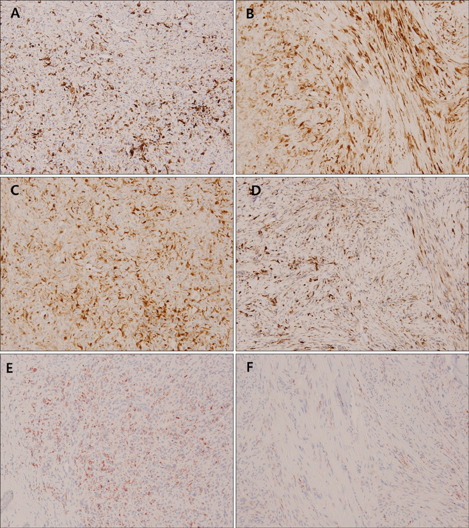
Fig. 1 Histological sections of dermatofibroma stained with antibodies against (A, B) leukocyte-specific protein 1 (LSP1), (C, D) factor XIIIa (FXIIIa), and (E, F) CD68. Expression of LSP1 and FXIIIa was observed in both (A, C) cellular lesions and (B, D) fibrous lesions. In contrast, immunoreactivity of CD68 was stronger in (E) cellular lesions than (F) fibrous lesions (A~F: ×200).

Fig. 2 Histological sections of dermatofibroma and normal skin stained with antibodies to (A, C) factor XIIIa (FXIIIa) and (B, D) leukocyte-specific protein 1 (LSP1). The expression patterns of (A) FXIIIa and (B) LSP1 were different in the same specimen of dermatofibroma. (C) FXIIIa-positive cells could be observed sporadically in the normal dermis, whereas (D) LSP1-positive cells could not be observed except for in a few lymphocytes in the normal dermis (A, B: ×40, C, D: ×100).
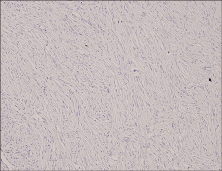
Fig. 3 Histological sections of dermatofibrosarcoma protuberans stained with leukocyte-specific protein 1 (LSP1) antibody. LSP1 staining in the lesion was negative (×200).
Reference
-
1. Song Y, Sakamoto F, Ito M. Characterization of factor XIIIa+ dendritic cells in dermatofibroma: Immunohistochemical, electron and immunoelectron microscopical observations. J Dermatol Sci. 2005; 39:89–96.
Article2. Horenstein MG, Prieto VG, Nuckols JD, Burchette JL, Shea CR. Indeterminate fibrohistiocytic lesions of the skin: is there a spectrum between dermatofibroma and dermatofibrosarcoma protuberans. Am J Surg Pathol. 2000; 24:996–1003.3. Yan X, Takahara M, Xie L, Tu Y, Furue M. Cathepsin K expression: a useful marker for the differential diagnosis of dermatofibroma and dermatofibrosarcoma protuberans. Histopathology. 2010; 57:486–488.
Article4. Goldblum JR, Tuthill RJ. CD34 and factor-XIIIa immunoreactivity in dermatofibrosarcoma protuberans and dermatofibroma. Am J Dermatopathol. 1997; 19:147–153.
Article5. Kuroda K, Tajima S. Proliferation of HSP47-positive skin fibroblasts in dermatofibroma. J Cutan Pathol. 2008; 35:21–26.
Article6. Cerio R, Spaull J, Jones EW. Histiocytoma cutis: a tumour of dermal dendrocytes (dermal dendrocytoma). Br J Dermatol. 1989; 120:197–206.
Article7. Bellini A, Mattoli S. The role of the fibrocyte, a bone marrow-derived mesenchymal progenitor, in reactive and reparative fibroses. Lab Invest. 2007; 87:858–870.
Article8. Aiba S, Tagami H. Phorbol 12-myristate 13-acetate can transform monocyte-derived dendritic cells to different cell types similar to those found in dermatofibroma. A possible in vitro model of the histogenesis of dermatofibroma. J Cutan Pathol. 1998; 25:65–71.
Article9. Yang L, Scott PG, Dodd C, Medina A, Jiao H, Shankowsky HA, et al. Identification of fibrocytes in postburn hypertrophic scar. Wound Repair Regen. 2005; 13:398–404.
Article10. Bucala R, Spiegel LA, Chesney J, Hogan M, Cerami A. Circulating fibrocytes define a new leukocyte subpopulation that mediates tissue repair. Mol Med. 1994; 1:71–81.
Article11. Blakaj A, Bucala R. Fibrocytes in health and disease. Fibrogenesis Tissue Repair. 2012; 5:Suppl 1. S6.
Article12. Jongstra-Bilen J, Misener VL, Wang C, Ginzberg H, Auerbach A, Joyner AL, et al. LSP1 modulates leukocyte populations in resting and inflamed peritoneum. Blood. 2000; 96:1827–1835.
Article13. Kanitakis J, Schmitt D, Thivolet J. Immunohistologic study of cellular populations of histiocytofibromas ("dermatofibromas"). J Cutan Pathol. 1984; 11:88–94.
Article14. Nestle FO, Nickoloff BJ, Burg G. Dermatofibroma: an abortive immunoreactive process mediated by dermal dendritic cells. Dermatology. 1995; 190:265–268.
Article15. Kuroda K, Tsukifuji R, Shinkai H. Increased expression of heat-shock protein 47 is associated with overproduction of type I procollagen in systemic sclerosis skin fibroblasts. J Invest Dermatol. 1998; 111:1023–1028.
Article16. Kubo M, Ihn H, Yamane K, Tamaki K. The expression levels and the differential expression of transforming growth factor-beta receptors in dermatofibroma and dermatofibrosarcoma protuberans. Br J Dermatol. 2006; 154:919–925.
Article17. Kim HJ, Lee JY, Kim SH, Seo YJ, Lee JH, Park JK, et al. Stromelysin-3 expression in the differential diagnosis of dermatofibroma and dermatofibrosarcoma protuberans: comparison with factor XIIIa and CD34. Br J Dermatol. 2007; 157:319–324.
Article18. Kahn HJ, Fekete E, From L. Tenascin differentiates dermatofibroma from dermatofibrosarcoma protuberans: comparison with CD34 and factor XIIIa. Hum Pathol. 2001; 32:50–56.
Article19. Sachdev R, Sundram U. Expression of CD163 in dermatofibroma, cellular fibrous histiocytoma, and dermatofibrosarcoma protuberans: comparison with CD68, CD34, and Factor XIIIa. J Cutan Pathol. 2006; 33:353–360.
Article
- Full Text Links
-
- Actions
-
Cited
- CITED
-
- Close
- Share
-
- Similar articles
-
- Analysis of Bone Marrow Micrometastasis using RT-PCR in Patients with Small Cell Lung Carcinoma
- A Case of Dermatofibroma with Granular Cells
- Macrophage/dendritic Cell Marker Staining Characteristics of Langerhans cell Granulomatosis(Histiocytosis X)
- Immunohistochemical Studies of bc1-2 Protein in a Skin Tumor of Neuroectodermal Origin
- Two Cases of Dermatofibroma with Atrophic Features
