Transcriptional Analysis of Nod-Like Receptors in a Mouse Model of Experimental Autoimmune Uveitis
- Affiliations
-
- 1Department of Ophthalmology, Seoul National University Hospital, Seoul National University College of Medicine, Seoul, Korea. jooyounoh77@gmail.com
- 2Department of Ophthalmology, Seoul Metropolitan Government Seoul National University Boramae Medical Center, Seoul, Korea.
- 3Laboratory of Ocular Regenerative Medicine and Immunology, Seoul Artificial Eye Center, Seoul National University Hospital Biomedical Research Institute, Seoul, Korea.
- KMID: 2216160
- DOI: http://doi.org/10.3341/jkos.2015.56.1.99
Abstract
- PURPOSE
To evaluate the transcription pattern of Nod-like receptors (NLRs), the intracellular sensors, to detect danger signals in murine eyes with experimental autoimmune uveitis (EAU).
METHODS
EAU was induced in B6 (C57BL/6) mice by subcutaneous injection of human interphotoreceptor retinoid binding protein and intraperitoneal injection of pertussis toxin. At 1, 2, and 3 weeks post-immunization, the eyeballs were extracted and subjected to histological and molecular assays using real-time reverse transcription polymerase chain reaction.
RESULTS
The levels of nucleotide-binding oligomerization domain, Leucine rich Repeat and Pyrin domain 1 (NLRP1), NLRP3, nucleotide-binding oligomerization domain-containing protein 1 (NOD1), and NOD2 transcripts were increased at 2 weeks and gradually reduced thereafter. Notably, NLRP3 showed the highest expression in the eyes with EAU. Similarly, the transcript level of pro-inflammatory cytokine, interleukin-1beta, increased and reached a peak at 2 weeks post-immunization. The retinal structure was severely damaged by inflammation at 3 weeks post-immunization.
CONCLUSIONS
Among NLRs, NLRP3 may induce inflammation in eyes after EAU immunization.
MeSH Terms
Figure
-
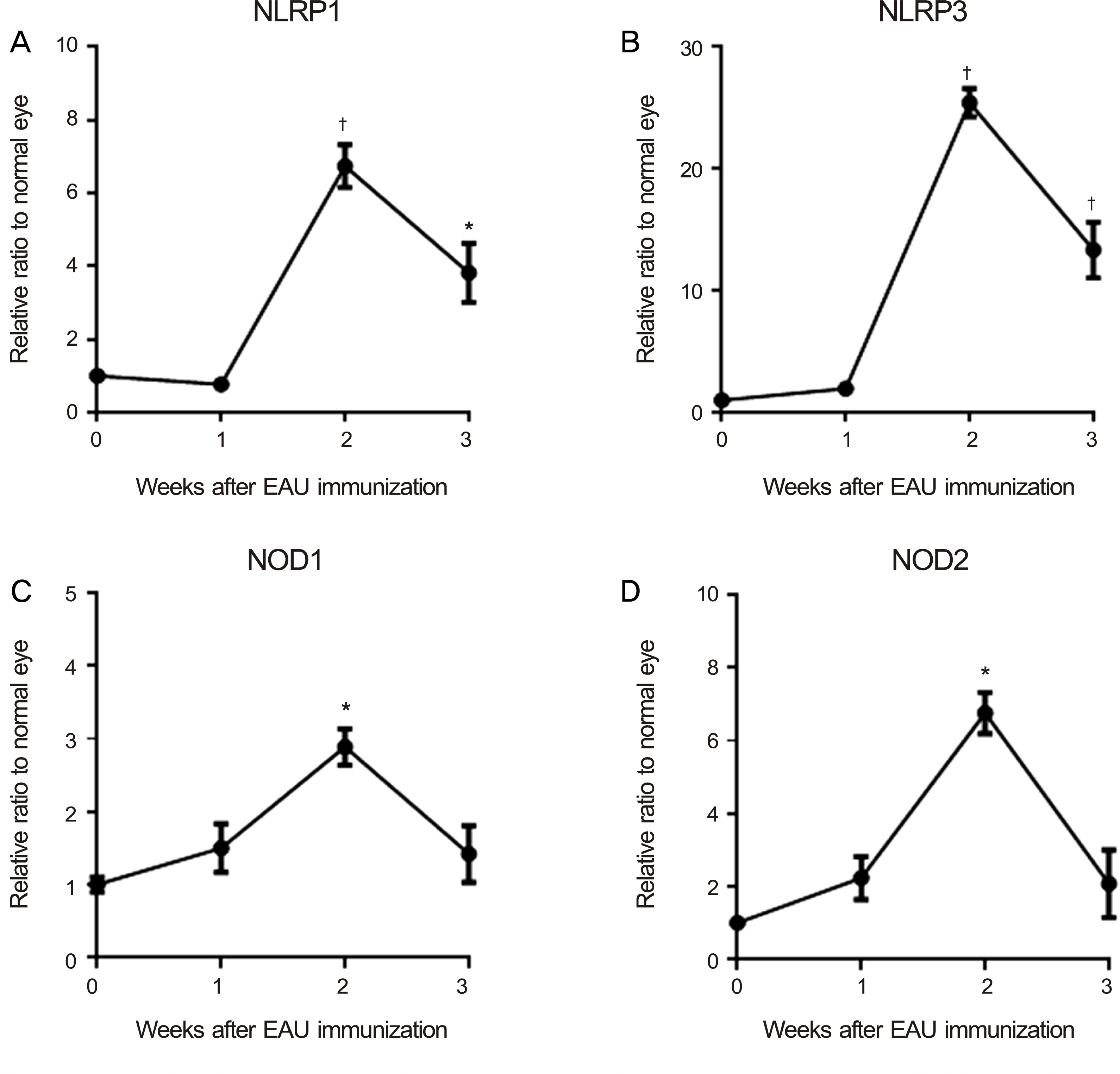
Figure 1. Assay for Nod-like receptors in the eyes after experimental autoimmune uveitis (EAU) immunization. Real-time RT-PCR analysis showed that the levels of NLRP1, NLRP3, NOD1, and NOD2 transcripts increased in the eyes after EAU immunization with a peak at the post-immunization 2 weeks, and gradually decreased until 3 weeks. (A) Time course of NLRP1 expression in the eye. (B) Time course of NLRP3 expression in the eye. (C) Time course of NOD1 expression in the eye. (D) Time course of NOD2 expression in the eye. Data are presented in mean ± SEM. N = 5 in each group. RT-PCR = reverse transcription polymerase chain reaction; NLRP = nucleotide-binding oligomerization domain, Leucine rich Repeat and Pyrin domain; NOD = nucleo-tide-binding oligomerization domain-containing protein; SEM = standard error of the mean. * p < 0.01; † p < 0.001.
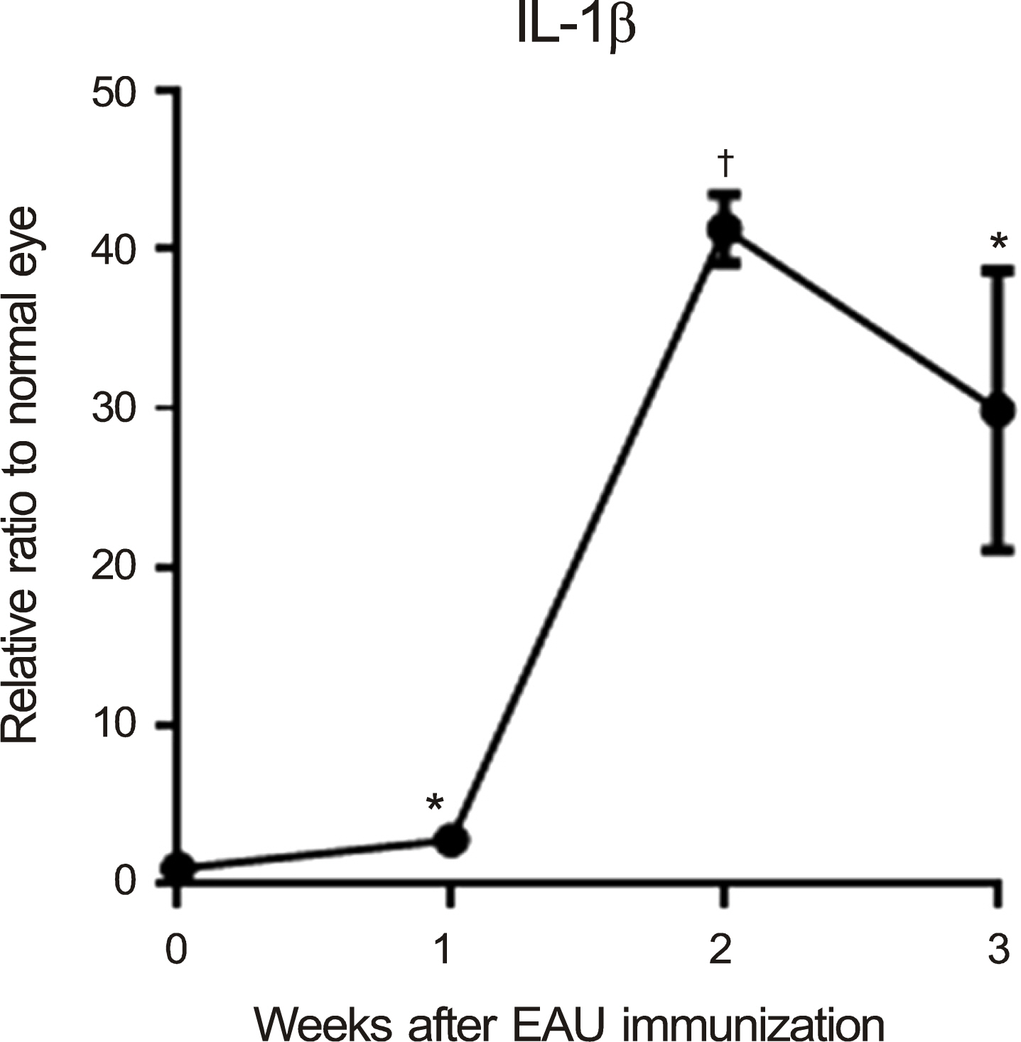
Figure 2. Assay for the pro-inflammatory cytokine interleukin (IL)-1β in the eyes after experimental autoimmune uveitis (EAU) immunization. Real-time RT-PCR analysis showed that the level of IL-1β transcript were gradually increased and reached at peak at the post-immunization 2 weeks after EAU immunization. Data are presented in mean ± SEM. N = 5 in each group. RT-PCR = reverse transcription polymerase chain reaction; SEM = standard error of the mean. * p < 0.05; † p < 0.001.
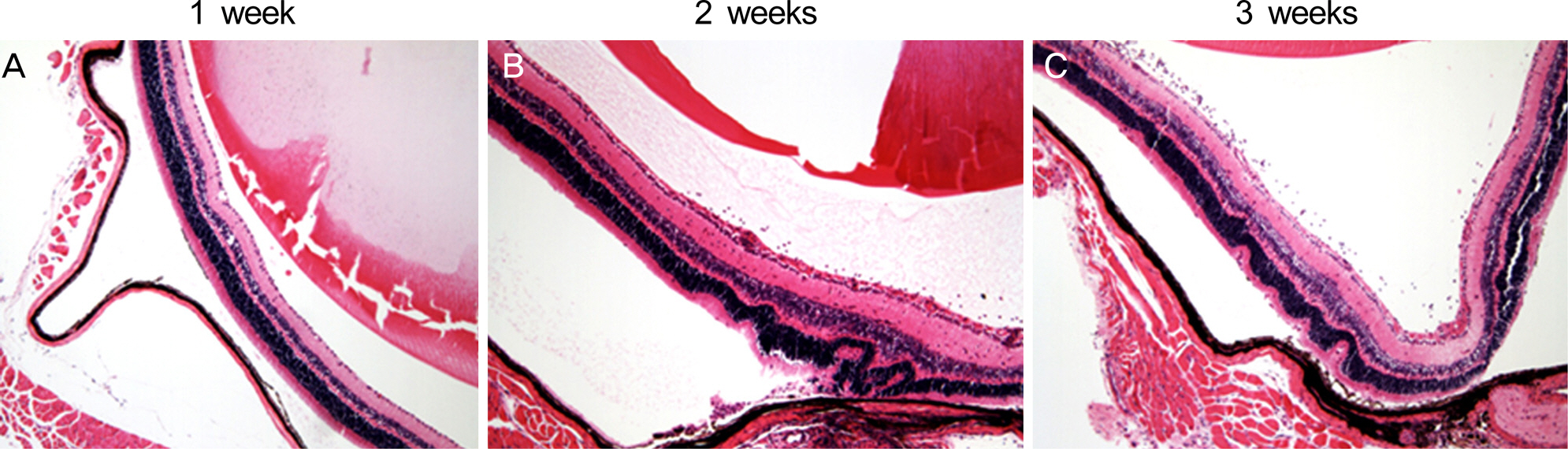
Figure 3. Histological findings of the eye. Hematoxylin-eosin staining of the eye showed that the retinal structure including the pho-toreceptor layer was severely disorganized with massive infiltration of inflammatory cells in the vitreous cavity and in the retina of BSS-treated experimental autoimmune uveitis mice on day 21 (Original magnification x100, Hematoxylin-eosin staining). (A) Hematoxylin-eosin staining of the eye at one week after EAU induction. (B) Hematoxylin-eosin staining of the eye at two weeks after EAU induction. (C) Hematoxylin-eosin staining of the eye at three weeks after EAU induction. BSS = balanced salt solution.
Reference
-
References
1. Chang JH, Wakefield D. Uveitis: a global perspective. Ocul Immunol Inflamm. 2002; 10:263–79.
Article2. Caspi RR. A look at autoimmunity and inflammation in the eye. J Clin Invest. 2010; 120:3073–83.
Article3. Martinon F, Tschopp J. NLRs join TLRs as innate sensors of pathogens. Trends Immunol. 2005; 26:447–54.
Article4. Ogura Y, Sutterwala FS, Flavell RA. The inflammasome: first line of the immune response to cell stress. Cell. 2006; 126:659–62.
Article5. Caspi RR. Experimental autoimmune uveoretinitis in the rat and mouse. Curr Protoc Immunol. 2003; Chapter 15:Unit 15.6.
Article6. Gritz DC, Wong IG. Incidence and prevalence of uveitis in Northern California; the Northern California Epidemiology of Uveitis Study. Ophthalmology. 2004; 111:491–500. discussion 500.
Article7. LeHoang P. The gold standard of noninfectious uveitis: corti- costeroids. Dev Ophthalmol. 2012; 51:7–28.8. Willermain F, Rosenbaum JT, Bodaghi B. . Interplay between innate and adaptive immunity in the development of non-infectious uveitis. Prog Retin Eye Res. 2012; 31:182–94.
Article9. Martinon F, Mayor A, Tschopp J. The inflammasomes: guardians of the body. Annu Rev Immunol. 2009; 27:229–65.
Article10. Strober W, Murray PJ, Kitani A, Watanabe T. Signalling pathways and molecular interactions of NOD1 and NOD2. Nat Rev Immunol. 2006; 6:9–20.
Article11. Vandanmagsar B, Youm YH, Ravussin A. . The NLRP3 inflammasome instigates obesity-induced inflammation and insulin resistance. Nat Med. 2011; 17:179–88.
Article12. Rosenzweig HL, Planck SR, Rosenbaum JT. NLRs in immune privileged sites. Curr Opin Pharmacol. 2011; 11:423–8.
Article13. Franchi L, Warner N, Viani K, Nuñez G. Function of Nod-like receptors in microbial recognition and host defense. Immunol Rev. 2009; 227:106–28.
Article14. Chen M, Wang H, Chen W, Meng G. Regulation of adaptive immunity by the NLRP3 inflammasome. Int Immunopharmacol. 2011; 11:549–54.
Article
- Full Text Links
-
- Actions
-
Cited
- CITED
-
- Close
- Share
-
- Similar articles
-
- Effects of Cyclosporin A on the Recurrence in Experimental Autoimmune Anterior Uveitis
- Defects in the differentiation and function of bone marrow-derived dendritic cells in non-obese diabetic mice
- Blood-retina barrier dysfunction in experimental autoimmune uveitis: the pathogenesis and therapeutic targets
- Expression of Cell Surface Receptors on Human Glioblastoma Xenograft Model in NOD/SCID Mouse
- Experimental Autoimmune Uveitis induced by Bovine Iris and Ciliary body in Lewis Rat
