Evaluation of Glaucomatous Damage in the Fellow Eyes of Patients With Unilateral Retinal Vein Occlusion
- Affiliations
-
- 1Department of Ophthalmolgy, University of Ulsan, Asan Medical Center, Seoul, Korea. mskook@amc.seoul.kr
- 2Han-Gil Eye Hospital, Incheon, Korea.
- 3Cheil Eye Hospital, Daegu, Korea.
- 4Hanyang University GURI Hospital, Guri-si, Gyeonggi-do, Korea.
- KMID: 2211906
- DOI: http://doi.org/10.3341/jkos.2009.50.1.120
Abstract
- PURPOSE
To investigate the visual field (VF) and retinal nerve fiber layer (RNFL) status of the fellow eyes in patients with unilateral retinal vein occlusion (RVO).
METHODS
Fifty patients with unilateral RVO and 35 normal control subjects wereconsecutively recruited. Humphrey VF parameters and RNFL status using scanning laser polarimetry with variable corneal compensation (GDx-VCC) were compared between the fellow eyes of the patients with unilateral RVO and control eyes. We also assessed the risk factors for the development of glaucomatous damage in the fellow eyes of unilateral RVO patients.
RESULTS
Twelve fellow eyes out of 50 patients with unilateral RVO showed glaucomatous VF and RNFL changes assessed by GDx-VCC. VF indices and RNFL thickness parameters in the study group were significantly lower than those in the control group (p<0.05). Increased age and vertical cup-to-disc ratio were significantly associated with severity of VF and RNFL damage in the fellow eye of unilateral RVO patients (p<0.05).
CONCLUSIONS
The fellow eyes in patients with unilateral RVO showed significantly worse VF indices and lower RNFL thickness than normal control eyes. The glaucomatous change should be carefully monitored in the fellow eyes of unilateral RVO patients.
Keyword
MeSH Terms
Figure
-
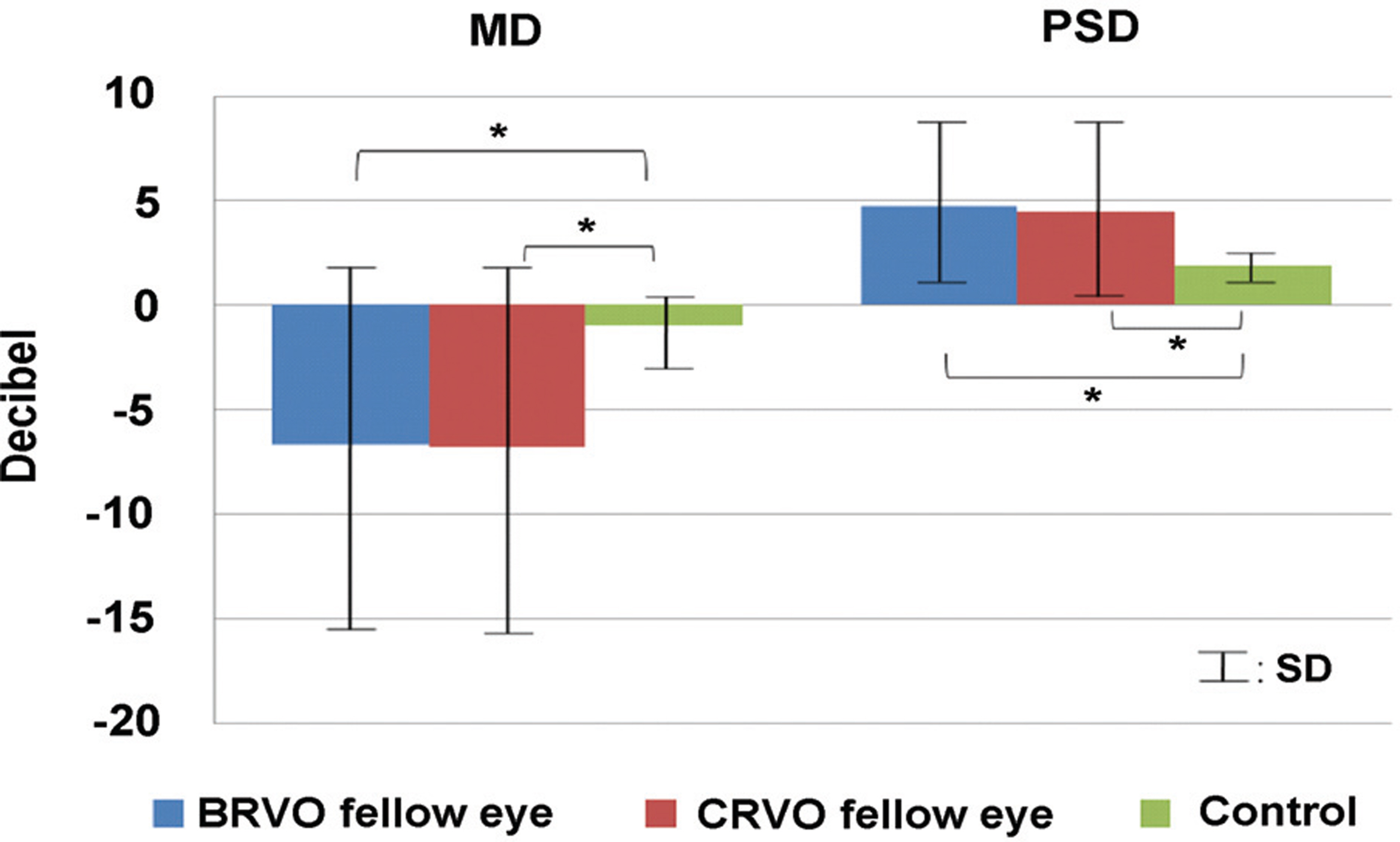
Figure 1. Comparison of visual field indices of the fellow eyes of branch retinal vein occlusion (BRVO) patients, the fellow eyes of central retinal vein occlusion (CRVO) patients and controls; MD=mean deviation; PSD=pattern standard deviation; BRVO=branch retinal vein occlusion; CRVO=central retinal vein * Statistical significance was found with occlusion; unpaired t-test (p<0.001).
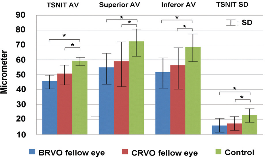
Figure 2. Global and sectoral RNFL retardation values based on GDx-VCC of the fellow eyes of branch retinal vein occlusion (BRVO) patients, the fellow eyes of central retinal vein occlusion (CRVO) patients and controls; TSNIT=temporal, superior, nasal, inferior and temporal AV=average; SD=standard deviation; BRVO= branch retinal vein occlusion; CRVO=central retinal vein occlusion; * Statistical significance was found with unpaired t-test (p<0.001).
Cited by 1 articles
-
Lamina Cribrosa Thickness in the Fellow Eyes of Patients with Unilateral Retinal Vein Occlusion
Yong Il Kim, Tea Yoon Lee, Kyoo Won Lee, Jin Seon Kim
J Korean Ophthalmol Soc. 2015;56(11):1736-1741. doi: 10.3341/jkos.2015.56.11.1736.
Reference
-
References
1. Krakau CE. Disk hemorrhages and retinal vein occlusions in glaucoma. Surv Ophthalmol. 1994; 38:S18–21.
Article2. Luntz MH, Schenker HI. Retinal vascular accidents in glaucoma and ocular hypertension. Surv Ophthalmol. 1980; 25:163–7.
Article3. Sonnsjö B, Krakau CE. Arguments for a vascular glaucoma etiology. Acta Ophthalmol. 1993; 71:433–44.
Article4. Rath EZ, Frank RN, Shin DH, Kim C. Risk factors for retinal vein occlusions. A case-control study. Ophthalmology. 1992; 99:509–14.5. The Eye Disease Case-Control Study Group Risk factors for branch retinal vein occlusion. Am J Ophthalmol. 1993; 116:286–96.6. The Eye Disease Case-Control Study Group Risk factors for central retinal vein occlusion. Arch Ophthalmol. 1996; 114:545–54.7. Kim SJ, Park KH. Four cases of normal-tension glaucoma with disk hemorrhage combined with BRVO in the contralateral eye. Am J Ophthalmol. 2004; 137:357–9.8. Hood DC, Thienprasiddhi P, Greenstein VC. . Detecting early to mild glaucomatous damage: a comparison of the multifocal VEP and automated perimetry. Invest Ophthalmol Vis Sci. 2004; 45:492–8.
Article9. Reus NJ, de Graaf M, Lemij HG. Accuracy of GDx VCC, HRT I, and clinical assessment of stereoscopic optic nerve head photographs for diagnosing glaucoma. Br J Ophthalmol. 2007; 91:313–8.
Article10. Oh SW, Ahn CS, Rhym MH. A clinical analysis on 186 cases of glaucoma. J Korean Ophthalmol Soc. 1970; 11:17–20.11. Shin SG, Ahn JH, Rho SH. A clinical analysis on 456 cases of glaucoma among outpatients during 5 years. J Korean Ophthalmol Soc. 1987; 28:1021–6.12. Song MS, Kim DG, Kim HJ. Clinical study on glaucomatous patients. J Korean Ophthalmol Soc. 1989; 30:755–9.13. Song KH, Jin KH, Kim JM. Clincal data on glaucoma patients. J Korean Ophthalmol Soc. 1990; 31:1179–83.14. Hwang IC, Jeong SK, Yang KJ. A clinical study on glaucoma. J Korean Ophthalmol Soc. 1992; 33:394–400.15. Lee CK, Cho YJ, Hong YJ. Distribution of glaucoma in out-patient clinic. J Korean Ophthalmol Soc. 1995; 36:1020–7.16. Lee CH, Jin GH, Kim DM. Clinical analysis on glaucoma. J Korean Ophthalmol Soc. 1998; 39:362–8.17. Bonovas S, Peponis V, Filioussi K. Diabetes mellitus as a risk factor for primary open-angle glaucoma: a meta-analysis. Diabet Med. 2004; 21:609–14.
Article18. Ellis JD, Evans JM, Ruta DA. . Glaucoma incidence in an unselected cohort of diabetic patients: is diabetes mellitus a risk factor for glaucoma? DARTS/MEMO collaboration: Diabetes Audit and Research in Tayside Study. Medicines Monitoring Unit. Br J Ophthalmol. 2000; 84:1218–24.19. Pasquale LR, Kang JH, Manson JE. . Prospective study of type 2 diabetes mellitus and risk of primary open-angle glaucoma in women. Ophthalmology. 2006; 113:1081–6.
Article20. Gordon MO, Beiser JA, Brandt JD. . The Ocular Hypertension Treatment Study: baseline factors that predict the onset of primary open-angle glaucoma. Arch Ophthalmol. 2002; 120:714–20.21. Hulsman CA, Vingerling JR, Hofman A. . Blood pressure, arterial stiffness, and open-angle glaucoma: the Rotterdam study. Arch Ophthalmol. 2007; 125:805–12.22. Hayreh SS, Zimmerman MB, Beri M, Podhajsky P. Intraocular pressure abnormalities associated with central and hemicentral retinal vein occlusion. Ophthalmology. 2004; 111:133–41.
Article23. Verhoeff FH. The effect of chronic glaucoma on the central retinal vessels. Arch Ophthalmol. 1913; 42:145–52.24. Moore RF. Retinal venous thrombosis: a clinical study of sixty-two cases followed over many years. Br J Ophthalmol. 1924; 8:S1–90.25. Duke-Elder S. Textbook of ophthalmology. Vol. 3. London: Kimpton;1936. p. 2584–5.
Article26. Becker B, Post LT Jr. Retinal vein occlusion. Clinical and experimental observations. Am J Ophthalmol. 1951; 34:677–86.27. Soni KG, Woodhouse DF. Retinal vascular occlusion as a presenting feature of glaucoma simplex. Br J Ophthalmol. 1971; 55:192–5.
Article28. Hitchings RA, Spaeth GL. Chronic retinal vein occlusion in glaucoma. Br J Ophthalmol. 1976; 60:694–9.
Article29. Grunwald JE, Riva CE, Stone RA. . Retinal autoregulation in open-angle glaucoma. Ophthalmology. 1984; 91:1690–4.
Article30. Pillunat LE, Stodtmeister R, Wilmanns I, Christ T. Auto-regulation of ocular blood flow during changes in intraocular pressure. Preliminary results. Graefes Arch Clin Exp Ophthal-mol. 1985; 223:219–23.31. Hayreh SS. Role of nocturnal arterial hypotension in the development of ocular manifestations of systemic arterial hypertension. Curr Opin Ophthalmol. 1999; 10:474–82.
Article32. Hayreh SS, Zimmerman MB, Podhajsky P, Alward WL. Nocturnal arterial hypotension and its role in optic nerve head and ocular ischemic disorders. Am J Ophthalmol. 1994; 117:603–24.
Article33. de Waard V, Arkenbout EK. Vos M. . TR3 nuclear orphan receptor prevents cyclic stretch-induced proliferation of venous smooth muscle cells. Am J Pathol. 2006; 168:2027–35.
Article34. Vikas Chopra, Rohit Varma. Brian A. . Type 2 diabetes mellitus and the risk of open-angle glaucoma the Los Angeles Latino Eye Study. Ophthalmology. 2008; 115:227–32.35. Sofi F, Mannini L, Marcucci R. . Role of haemorheological factors in patients with retinal vein occlusion. Thromb Haemost. 2007; 98:1215–9.
Article36. Leoncini G, Bruzzese D, Signorello MG. . Platelet activation by collagen is increased in retinal vein occlusion. Thromb Haemost. 2007; 97:218–27.
Article37. Klaver JHJ, Greve EL, Goslinga H. . Blood and plasma viscosity measurements in patients with glaucoma. Br J Ophthalmol. 1985; 69:765.
Article38. Drance SM, Sweeney VP, Morgan RW. . Studies of factors involved in the production of low tension glaucoma. Arch Ophthalmol. 1973; 89:457.
Article39. Crichton A, Drance SM, Douglas GR, Schulzer M. Unequal intraocular pressure and its relation to asymmetric visual field defects in low tension glaucoma. Ophthalmology. 1989; 96:1312–4.40. Choi J, Kim KH, Jeong J. . Circadian fluctuation of mean ocular perfusion pressure is a consistent risk factor for normal- tension glaucoma. Invest Ophthalmol Vis Sci. 2007; 48:104–11.41. Plange N, Remky A, Arend O. Absolute filling defects of the optic disc in fluorescein angiograms inglaucoma--a retro- spective clinical study. Klin Monatsbl Augenheilkd. 2001; 218:214–21.42. Pollack A, Dottan S, Oliver M. The fellow eye in retinal vein occlusive disease. Ophthalmology. 1989; 96:842–5.
Article43. Beaumont PE, Kang HK. Cup-to-disc ratio, intraocular pressure, and primary open-angle glaucoma in retinal venous occlusion. Ophthalmology. 2002; 109:282–6.
- Full Text Links
-
- Actions
-
Cited
- CITED
-
- Close
- Share
-
- Similar articles
-
- Lamina Cribrosa Thickness in the Fellow Eyes of Patients with Unilateral Retinal Vein Occlusion
- Comparison between Glaucomatous and Non-glaucomatous Eyes with Unilateral Retinal Vein Occlusion in the Fellow Eye
- Clinical observation of the bilateral branch vein occlusion
- Comparison of Inner Retinal Thickness between the Fellow Eyes of Unilateral Branch Retinal Vein Occlusion and Normal Control
- Diurnal Fluctuation of Ocular Perfusion Pressure in the Fellow Eyes of Branch Retinal Vein Occlusion
