Korean J Ophthalmol.
2013 Dec;27(6):440-445. 10.3341/kjo.2013.27.6.440.
Comparison between Glaucomatous and Non-glaucomatous Eyes with Unilateral Retinal Vein Occlusion in the Fellow Eye
- Affiliations
-
- 1Department of Ophthalmology, Asan Medical Center, University of Ulsan College of Medicine, Seoul, Korea. sungeye@gmail.com
- KMID: 1792080
- DOI: http://doi.org/10.3341/kjo.2013.27.6.440
Abstract
- PURPOSE
To evaluate and compare the clinical and angiographic characteristics of retinal vein occlusion (RVO) in glaucomatous and non-glaucomatous eyes with unilateral RVO in the fellow eye.
METHODS
Twenty-one glaucomatous eyes (GL group) and 25 age-matched non-glaucomatous eyes (non-GL group) with unilateral RVO in the fellow eye were included in this study. Fluorescein angiographic images were assessed in both groups by 3 retina specialists in order to determine the RVO occlusion site. The occlusion site was divided into 2 types: arteriovenous (AV)-crossing and non-AV-crossing (optic cup or optic nerve sited). The clinical characteristics and prevalence of AV-crossing and non-AV-crossing RVO were compared between the 2 groups.
RESULTS
The mean baseline intraocular pressures of the RVO eye and the fellow eye did not differ between the 2 groups (RVO eye: 14.3 +/- 2.5 mmHg [non-GL group], 15.5 +/- 3.9 mmHg [GL group], p = 0.217; fellow eye: 14.4 +/- 2.5 mmHg [non-GL group], 15.7 +/- 3.7 mmHg [GL group], p = 0.148). The prevalence of systemic disease did not differ between the 2 groups (e.g., diabetes mellitus and hypertension, p = 0.802 and 0.873, respectively). AV-crossing RVO was significantly more frequent in the non-GL group (19 eyes; 76%) than in the GL group (4 eyes, 19%, p < 0.001).
CONCLUSIONS
Non-AV-crossing RVO, i.e., optic cup- or optic nerve-sited RVO, is more frequently associated with glaucomatous changes in the fellow eye. Therefore, this type of RVO should be monitored more carefully for indications of glaucoma in the fellow eye.
Keyword
MeSH Terms
Figure
-

Fig. 1 Clinical case of non-glaucomatous eye with unilateral retinal vein occlusion (RVO) in the fellow eye. This patient had arteriovenous crossing RVO in the right eye (A) and non-glaucomatous eye in the left (B).
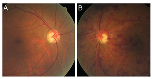
Fig. 2 Clinical case of glaucomatous eye with unilateral retinal vein occlusion (RVO) in the fellow eye. This patient had glaucomatous eye in the right (A) and non-arteriovenous-crossing type RVO (B) in the left eye.
Cited by 1 articles
-
Lamina Cribrosa Thickness in the Fellow Eyes of Patients with Unilateral Retinal Vein Occlusion
Yong Il Kim, Tea Yoon Lee, Kyoo Won Lee, Jin Seon Kim
J Korean Ophthalmol Soc. 2015;56(11):1736-1741. doi: 10.3341/jkos.2015.56.11.1736.
Reference
-
1. The Eye Disease Case-Control Study Group. Risk factors for central retinal vein occlusion. Arch Ophthalmol. 1996; 114:545–554.2. Soni KG, Woodhouse DF. Retinal vascular occlusion as a presenting feature of glaucoma simplex. Br J Ophthalmol. 1971; 55:192–195.3. Kim MJ, Woo SJ, Park KH, Kim TW. Retinal nerve fiber layer thickness is decreased in the fellow eyes of patients with unilateral retinal vein occlusion. Ophthalmology. 2011; 118:706–710.4. Klein BE, Meuer SM, Knudtson MD, Klein R. The relationship of optic disk cupping to retinal vein occlusion: the Beaver Dam Eye Study. Am J Ophthalmol. 2006; 141:859–862.5. Mitchell P, Smith W, Chang A. Prevalence and associations of retinal vein occlusion in Australia. The Blue Mountains Eye Study. Arch Ophthalmol. 1996; 114:1243–1247.6. Beaumont PE, Kang HK. Clinical characteristics of retinal venous occlusions occurring at different sites. Br J Ophthalmol. 2002; 86:572–580.7. Beaumont P, Goldberg I, Hollows FC. Optic cup vein occlusion: description of a new entity. Trans Ophthalmol Soc N Z. 1976; 28:115–117.8. Kim CS, Seong GJ, Lee NH, et al. Prevalence of primary open-angle glaucoma in central South Korea the Namil study. Ophthalmology. 2011; 118:1024–1030.9. Iwase A, Suzuki Y, Araie M, et al. The prevalence of primary open-angle glaucoma in Japanese: the Tajimi Study. Ophthalmology. 2004; 111:1641–1648.10. Beaumont PE, Kang HK. Pattern of vascular nonperfusion in retinal venous occlusions occurring within the optic nerve with and without optic nerve head swelling. Arch Ophthalmol. 2000; 118:1357–1363.11. Lim LL, Cheung N, Wang JJ, et al. Prevalence and risk factors of retinal vein occlusion in an Asian population. Br J Ophthalmol. 2008; 92:1316–1319.12. Tan AG, Mitchell P, Burlutsky G, et al. Retinal vessel caliber and the long-term incidence of age-related cataract: the Blue Mountains Eye Study. Ophthalmology. 2008; 115:1693–1698.13. Cugati S, Wang JJ, Knudtson MD, et al. Retinal vein occlusion and vascular mortality: pooled data analysis of 2 population-based cohorts. Ophthalmology. 2007; 114:520–524.14. Jiang X, Varma R, Wu S, et al. Baseline risk factors that predict the development of open-angle glaucoma in a population: the Los Angeles Latino Eye Study. Ophthalmology. 2012; 119:2245–2253.15. Kim YJ, Yun SC, Na JH, et al. Glaucoma progression in eyes with a history of refractive corneal surgery. Invest Ophthalmol Vis Sci. 2012; 53:4485–4489.16. Leske MC, Heijl A, Hyman L, et al. Predictors of long-term progression in the early manifest glaucoma trial. Ophthalmology. 2007; 114:1965–1972.
- Full Text Links
-
- Actions
-
Cited
- CITED
-
- Close
- Share
-
- Similar articles
-
- Evaluation of Glaucomatous Damage in the Fellow Eyes of Patients With Unilateral Retinal Vein Occlusion
- Lamina Cribrosa Thickness in the Fellow Eyes of Patients with Unilateral Retinal Vein Occlusion
- Comparison of Inner Retinal Thickness between the Fellow Eyes of Unilateral Branch Retinal Vein Occlusion and Normal Control
- Glaucomatous Optic Nerve Damage in the Fellow Eyes of Acute Angle-Closure Glaucoma
- Diurnal Fluctuation of Ocular Perfusion Pressure in the Fellow Eyes of Branch Retinal Vein Occlusion
