Syringo-Subarachnoid-Peritoneal Shunt Using T-Tube for Treatment of Post-Traumatic Syringomyelia
- Affiliations
-
- 1Department of Neurosurgery, School of Medicine, Chungnam National University, Daejeon, Korea. swchoi@cnu.ac.kr
- 2Brain Research Institute, Chungnam National University, Daejeon, Korea.
- KMID: 2190526
- DOI: http://doi.org/10.3340/jkns.2012.52.1.58
Abstract
- Various surgical procedures for the treatment of post-traumatic syringomyelia have been introduced recently, but most surgical strategies have been unreliable. We introduce the concept and technique of a new shunting procedure, syringo-subarachnoid-peritoneal shunt. A 54-year-old patient presented to our hospital with a progressive impairment of motion and position sense on the right side. Sixteen years before this admission, he had been treated by decompressive laminectomy for a burst fracture of L1. On his recent admission, magnetic resonance (MR) imaging studies of the whole spine revealed the presence of a huge syrinx extending from the medulla to the L1 vertebral level. We performed a syringo-subarachnoid-peritoneal shunt, including insertion of a T-tube into the syrinx, subarachnoid space and peritoneal cavity. Clinical manifestations and radiological findings improved after the operation. The syringo-subarachnoid-peritoneal shunt has several advantages. First, fluid can communicate freely between the syrinx, the subarachnoid space, and the peritoneal cavity. Secondly, we can prevent shunt catheter from migrating because dural anchoring of the T-tube is easy. Finally, we can perform shunt revision easily, because only one arm of the T-tube is inserted into the intraspinal syringx cavity. We think that this procedure is the most beneficial method among the various shunting procedures.
Keyword
MeSH Terms
Figure
-

Fig. 1 Magnetic resonance images of the cervical (A) and thoracic spine (B) reveal the presence of a huge syrinx extending from the medulla to the L1 level.

Fig. 2 Schematic diagram showing the technique of syringo-subarachnoid-peritoneal shunt. The T-tube arms which have many side holes, are cut to the desired length and one arm is inserted into the syrinx (rostral direction), the other arm into subarachnoid space (caudal direction). CSF: cerebrospinal fluid.
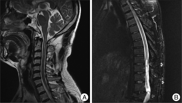
Fig. 3 Postoperative magnetic resonance images of cervical (A) and thoracic (B) at 27 months after the surgery show considerable reduction in the size of the syringomyelic cavity.
Cited by 1 articles
-
Treatment of Syringomyelia due to Chiari Type I Malformation with Syringo-Subarachnoid-Peritoneal Shunt
Akın Akakın, Baran Yılmaz, Murat Şakir Ekşi, Türker Kılıç
J Korean Neurosurg Soc. 2015;57(4):311-313. doi: 10.3340/jkns.2015.57.4.311.
Reference
-
1. Aghakhani N, Baussart B, David P, Lacroix C, Benoudiba F, Tadie M, et al. Surgical treatment of posttraumatic syringomyelia. Neurosurgery. 2010; 66:1120–1127. discussion 1127. PMID: 20495426.
Article2. Barbaro NM, Wilson CB, Gutin PH, Edwards MS. Surgical treatment of syringomyelia. Favorable results with syringoperitoneal shunting. J Neurosurg. 1984; 61:531–538. PMID: 6747690.3. Batzdorf U, Klekamp J, Johnson JP. A critical appraisal of syrinx cavity shunting procedures. J Neurosurg. 1998; 89:382–388. PMID: 9724111.
Article4. Brodbelt AR, Stoodley MA. Post-traumatic syringomyelia : a review. J Clin Neurosci. 2003; 10:401–408. PMID: 12852875.5. Cho KH, Iwasaki Y, Imamura H, Hida K, Abe H. Experimental model of posttraumatic syringomyelia : the role of adhesive arachnoiditis in syrinx formation. J Neurosurg. 1994; 80:133–139. PMID: 8270999.
Article6. Gardner WJ. Hydrodynamic factors in Dandy-Walker and Arnold-Chiari malformations. Childs Brain. 1977; 3:200–212. PMID: 891301.
Article7. Goel A, Desai K. Surgery for syringomyelia : an analysis based on 163 surgical cases. Acta Neurochir (Wien). 2000; 142:293–301. discussion 301-302. PMID: 10819260.8. Lee TT, Alameda GJ, Camilo E, Green BA. Surgical treatment of post-traumatic myelopathy associated with syringomyelia. Spine (Phila Pa 1976). 2001; 26:S119–S127. PMID: 11805618.
Article9. Lund-Johansen M, Wester K. Syringomyelia treated with a nonvalved syringoperitoneal shunt : a follow-up study. Neurosurgery. 1997; 41:858–864. discussion 864-865. PMID: 9316047.10. Perrouin-Verbe B, Lenne-Aurier K, Robert R, Auffray-Calvier E, Richard I, Mauduyt de la Grève I, et al. Post-traumatic syringomyelia and post-traumatic spinal canal stenosis : a direct relationship : review of 75 patients with a spinal cord injury. Spinal Cord. 1998; 36:137–143. PMID: 9495005.
Article11. Schaller B, Mindermann T, Gratzl O. Treatment of syringomyelia after posttraumatic paraparesis or tetraparesis. J Spinal Disord. 1999; 12:485–488. PMID: 10598990.
Article12. Schurch B, Wichmann W, Rossier AB. Post-traumatic syringomyelia (cystic myelopathy) : a prospective study of 449 patients with spinal cord injury. J Neurol Neurosurg Psychiatry. 1996; 60:61–67. PMID: 8558154.
Article13. Sgouros S, Williams B. A critical appraisal of drainage in syringomyelia. J Neurosurg. 1995; 82:1–10. PMID: 7815110.
Article14. Sgouros S, Williams B. Management and outcome of posttraumatic syringomyelia. J Neurosurg. 1996; 85:197–205. PMID: 8755746.
Article15. Vassilouthis J, Papandreou A, Anagnostaras S. Thecoperitoneal shunt for post-traumatic syringomyelia. J Neurol Neurosurg Psychiatry. 1994; 57:755–756. PMID: 8006663.
Article16. Vengsarkar US, Panchal VG, Tripathi PD, Patkar SV, Agarwal A, Doshi PK, et al. Percutaneous thecoperitoneal shunt for syringomyelia. Report of three cases. J Neurosurg. 1991; 74:827–831. PMID: 2013782.
- Full Text Links
-
- Actions
-
Cited
- CITED
-
- Close
- Share
-
- Similar articles
-
- Treatment of Syringomyelia due to Chiari Type I Malformation with Syringo-Subarachnoid-Peritoneal Shunt
- Evaluation of Syringo-Subarachnoid Shunt for Syringomyelia
- Syringo-Pleural Shunt for Failed Syringosubarachnoid Shunt in Posttraumatic Syringomyelia
- A Case of Syringomyelia Following Cured Tuberculous Meningitis
- Clinical Analysis of Post-traumatic Hydrocephalus
