Value of Coronary Calcium Score in Type 2 Diabetics
- Affiliations
-
- 1Department of Internal Medicine, College of Medicine, Yeungnam University, Korea.
- 2Department of Nuclear Medicine, College of Medicine, Yeungnam University, Korea.
- 3Ulsan Hospital, Korea.
- KMID: 2177602
- DOI: http://doi.org/10.4093/jkda.2006.30.4.303
Abstract
-
BACKGROUND: Cardiovascular disease including coronary heart disease (CHD) is the most common cause of morbidity and mortality in patients with diabetes. But traditional risk factor assessment is limited to predict CHD in asymptomatic high-risk individuals. In this study, relationship between coronary calcium score (CCS) and CHD was evaluated to determine value of coronary artery calcification detected by multi-slice spiral computed tomography to predict CHD in high risk asymptomatic patients with type 2 diabetes.
METHODS
127 patients were enrolled who admitted in Yeungnam University Hospital between December 2004 and May 2005. Standard cardiovascular risk factors and the CCS measured by multi-slice spiral computed tomography were assessed.
RESULTS
Enrolled subjects were consisted of 56 subjects with diabetes and 71 subjects without diabetes. The mean CCS was significantly greater in patients with diabetes than without diabetics (P < 0.01). In both groups, patients with higher CCS had higher prevalence of CHD (P < 0.05). In all subjects, LDL cholesterol levels and CCS were significantly associated in multi-variate analysis (P < 0.05). In patients without diabetes, age was only associated with presence of CHD (P < 0.05). CCS was only associated with CHD in patients with diabetes, even after adjusting for the effects of age, LDL cholesterol and CRP (P < 0.05).
CONCLUSION
Therefore, multi-slice spiral computed tomography can non-invasively and accurately detect coronary calcification. By detection of coronary artery calcification, it may be possible to predict coronary heart disease early in high-risk asymptomatic patients with type 2 diabetes.
MeSH Terms
Figure
-
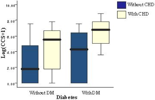
Fig. 1 Distribution of coronary calcium score in diabetics and non-diabetics. Total coronary calcium score was normalized by taking natural log of (1 + coronary calcium score). Diabetes group had higher coronary calcium score than non-diabetes group. Patients with significant coronary stenoses had significantly higher calcification scores than patients without stenoses in both the diabetic patients and non-diabetic patients.
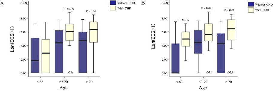
Fig. 2 Distribution of coronary calcium score stratified by age in patients with and without diabetes (A) or in diabetics with and without coronary heart disease(B). Because coronary artery calcium accumulates exponentially in advanced lesions and in older patients, we plotted the log-transformed calcification score after adjusting for patient age.
Reference
-
1. Haffner SM, Lehto S, Ronnemaa T, Pyorala K, Laakso M. Mortality from coronary heart disease in subjects with type 2 diabetes and in nondiabetic subjects with and without myocardial infarction. N Engl J Med. 1998. 339:229–234.2. Geiss LS. Diabetes in America. 1995. 2nd ed. Washington DC, U.S.: Govt Printing Office;233–257.3. Haffner SM. Coronary heart disease in patients with diabetes. N Engl J Med. 2000. 342:1040–1042.4. Gordon T, Castelli WP, Hjortland MC, Kannel WB, Dawber TR. Diabetes, blood lipids, and the role of obesity in coronary heart disease risk for women. Ann Intern Med. 1977. 87:393–397.5. Gordon T, Kannel WB. Multiple risk functions for predicting coronary heart disease: the concept, accuracy, and application. Am Heart J. 1982. 103:1031–1039.6. Kannel WB, McGee DL. Diabetes and glucose tolerance as risk factors for cardiovascular disease: the Framingham Study. Diabetes Care. 1979. 2:120–126.7. Budoff MJ. Atherosclerosis imaging and calcified plaque: coronary artery disease risk assessment. Prog Cardiovasc Dis. 2003. 46:135–148.8. Folsom AR, Evans GW, Carr JJ, Stillman AE. Association of traditional and nontraditional cardiovascular risk factors with coronary artery calcification. Angiology. 2004. 55:613–623.9. Greenland P, LaBree L, Azen SP, Doherty TM, Detrano RC. Coronary artery calcium score combined with Framingham score for risk prediction in asymptomatic individuals. JAMA. 2004. 291:210–215.10. Simons DB, Schwarz RS, Edwards WD, Sheedy PF, Breen JF, Rumberger JA. Noninvasive definition of anatomic coronary artery disease by ultrafast computed tomographic scanning: a quantitative pathologic comparison study. J Am Coll Cardiol. 1992. 20:1118–126.11. O'Rourke RA, Brundage BH, Froelicher VF, Greenland P, Grundy SM, Hachamovitch R, Pohost GM, Shaw LJ, Weintraub WS, Winters WL Jr, Forrester JS, Douglas PS, Faxon DP, Fisher JD, Gregoratos G, Hochman JS, Hutter AM Jr, Kaul S, Wolk MJ. American College of Cardiology/American Heart Association Expert Consensus document on electron-beam computed tomography for the diagnosis and prognosis of coronary artery disease. Circulation. 2000. 102:126–140.12. Schmermund A, Baumgart D, Gorge G, Seibel R, Gronemeyer D, Erbel R. Non-invasive visualization of coronary arteries with and without calcification by electron beam computed tomography. Herz. 1996. 21:118–126.13. Qu W, Le TT, Azen SP, Xiang M, Wong ND, Doherty TM, Detrano RC. Value of coronary artery calcium scanning by computed tomography for predicting coronary heart disease in diabetic subjects. Diabetes Care. 2003. 26:905–910.14. Frick M, Karakolcu F, Gaschnitzer H, Alber HF, Stoeger A, Obrist P, Friderich G, Weidinger F, Pachinger O, Schwarzacher SP. Calcium score as assessed by multi-slice computed tomography does not predict maximum plaque burden: an in vitro study. Heart. 2004. 90:1057–1058.15. Arad Y, Spadaro LA, Goodman K, Lledo-Perez A, Sherman S, Lerner G, Guerci AD. Predictive value of electron beam computed tomography of the coronary arteries. 19-month follow-up of 1173 asymptomatic subjects. Circulation. 1996. 93:1951–1953.16. Detrano R, Hsiai T, Wang S, Puentes G, Fallavollita J, Shields P, Stanford W, Wolfkiel C, Georgiou D, Budoff M, Reed J. Prognostic value of coronary calcification and angiographic stenoses in patients undergoing coronary angiography. J Am Coll Cardiol. 1996. 27:285–290.17. Secci A, Wong N, Tang W, Wang S, Doherty T, Detrano R. Electron beam computed tomographic coronary calcium as a predictor of coronary events: comparison of two protocols. Circulation. 1997. 96:1122–1129.18. Gerber TC, Kuzo RS, Karstaedt N, Lane GE, Morin RL, Sheedy PF 2nd, Safford RE, Blackshear JL, Pietan JH. Current results and new developments of coronary angiography with use of contrast-enhanced computed tomography of the heart. Mayo Clin Proc. 2002. 77:55–71.19. National Cholesterol Education Program (NCEP) Expert Panel on Detection, Evaluation, and Treatment of High Blood Cholesterol in Adults (Adult Treatment Panel III). Third Report of the National Cholesterol Education Program (NCEP) Expert Panel on Detection, Evaluation, and Treatment of High Blood Cholesterol in Adults (Adult Treatment Panel III) final report. Circulation. 2002. 106:3143–3421.20. Stone NJ, Bilek S, Rosenbaum S. Recent National Cholesterol Education Program Adult Treatment Panel III update: adjustments and options. Am J Cardiol. 2005. 96:53E–59E.21. American Diabetes Association. Standards of medical care in diabetes. Diabetes Care. 2005. 28:Suppl 1. S4–36.22. Chobanian AV, Bakris GL, Black HR, Cushman WC, Green LA, Izzo JL Jr, Jones DW, Materson BJ, Oparil S, Wright JT Jr, Roccella EJ. Joint National Committee on Prevention, Detection, Evaluation, and Treatment of High Blood Pressure. National Heart, Lung, and Blood Institute. National High Blood Pressure Education Program Coordinating Committee. Seventh report of the Joint National Committee on Prevention, Detection, Evaluation, and Treatment of High Blood Pressure. Hypertension. 2003. 42:1206–1252.23. Agatston AS, Janowitz WR, Hildner FJ, Zusmer NR, Viamonte M Jr, Detrano R. Quantification of coronary artery calcium using ultrafast computed tomography. J Am Coll Cardiol. 1990. 15:827–832.24. Kannel WB, Schatzkin A. Sudden death: lessons from subsets in population studies. J Am Coll Cardiol. 1985. 5:6 Suppl. 141B–149B.25. Grundy SM, Benjamin IJ, Bruke GL, Chait A, Eckel RH, Howard BV, Mitch W, Smith SC Jr, Sowers JR. Diabetes and cardiovascular disease: a statement for healthcare professionals from the American Heart Association. Circulation. 1999. 100:1134–1146.26. Hosoi M, Sato T, Yamagami K, Hasegawa T, Yamakita T, Miyamoto M, Yoshioka K, Yamamoto T, Ishii T, Tanaka S, Itoh A, Haze K, Fujii S. Impact of diabetes on coronary stenosis and coronary artery calcification detected by electron-beam computed tomography in symptomatic patients. Daibetes Care. 2002. 25:696–701.27. Rumberger JA, Brundage BH, Rader DJ, Kondos G. Electron beam computed tomographic coronary calcium scanning: a review and guidelines for use in asymptomatic persons. Mayo Clin Proc. 1999. 74:243–252.28. Schmermund A, Mohlenkamp S, Erbel R. Coronary artery calcium and its relationship to coronary disease. Cardiol Clin. 2003. 21:521–534.29. LaMonte MJ, FitzGerald SJ, Church TS, Barlow CE, Radford NB, Levine BD, Pippin JJ, Gibbons LW, Blair SN, Nichaman MZ. Coronary artery calcium score and coronary heart disease events in a large cohor of asymptomatic men and women. Am J Epidemiol. 2005. 162:421–429.30. Olson JC, Edmundowicz D, Becker DJ, Kuller LH, Orchard TJ. Coronary calcium in adults with type 1 diabetes: a stronger correlate of clinical coronary artery disease in men than in women. Diabetes. 2000. 49:1571–1578.31. Raggi P, Shaw LJ, Berman DS, Callister TQ. Prognostic value of coronary artery calcium screening in subjects with and without diabetes. J Am Coll Cardiol. 2004. 43:1663–1669.32. Rumberger JA, Sheedy PF, Breen JF, Schwarz RS. Electron beam computed tomographic coronary calcim score cutpoints and severity of associated angiographic lumen stenosis. J Am Coll Cardiol. 1997. 29:1542–1548.33. Arad Y, Spadaro LA, Goodman K, Newstein D, Guerci AD. Prediction of coronary events with electron beam computed tomography. J Am Coll Cardiol. 2000. 36:1253–1260.34. Revean PD, Sacks J. Investigators for the VADT: Coronary artery and abdominal aortic calcification are associated with cardiovascular disease in type 2 diabetes. Diabetologia. 2005. 48:379–385.35. Horiguchi J, Shen Y, Akiyama Y, Hirai N, Sasaki K, Ishifuro M, Nakanishi T, Ito K. Electron beam CT versus 16-MDCT on the variability of repeated coronary artery calcium measurements in a variable heart phantom. Am J Roentgenol. 2005. 185:995–1000.36. Knez A, Becker C, Becker A, Leber A, White C, Reiser M, Steinbeck G. Determination of coronary calcium with multi-slice spiral computed tomography: a comparative study with electron-beam CT. Int J Cardiovasc Imaging. 2002. 18:295–303.37. Stanford W, Thompson BH, Burns TL, Heery SD, Burr MC. Coronary artery calcium quantification at multi-detector row helical CT versus electron-beam CT. Radiology. 2004. 230:397–402.38. Hoffmann U, Moselewski F, Cury RC, Ferencik M, Jang IK, Diaz LJ, Abbara S, Brady TJ, Achenbach S. Predictive value of 16-slice multidetector spiral computed tomography to detect significant obstructive coronary artery disease in patients at high risk for coronary artery disease: patient-versus segment-based analysis. Circulation. 2004. 110:2638–2643.
- Full Text Links
-
- Actions
-
Cited
- CITED
-
- Close
- Share
-
- Similar articles
-
- The Role of Coronary Artery Calcium Score Study: for the Prevention and Reduction of Obstructive Coronary Arterial Disease
- The Relation of Coronary Artery Calcium Scores with Framingham Risk Scores
- Analysis of Aqueous Humor Calcium and Phosphate from Cataract Eyes with and without Diabetes Mellitus
- Which Individuals Could Benefit from Repeat Coronary Calcium Scans among Asymptomatic Korean Adults with a Baseline Coronary Artery Calcium Score of Zero?
- Relationship Between Serum Bilirubin Levels and Coronary Atherosclerosis in Patients with Type 2 Diabetes
