Complete biologic response to taxane based chemotherapy confirmed by [18F]FDG PET/CT and surgery in a cancer of unknown primary site
- Affiliations
-
- 1Department of Hematology-Oncology, Chonnam National University Medical School, Gwangju, Korea. ijchung@chonnam.ac.kr
- KMID: 2177516
- DOI: http://doi.org/10.3802/jgo.2012.23.1.65
Abstract
- Cancers of an unknown primary site are heterogenous with respect to their clinical and pathologic features. They are generally very aggressive, but specific favorable subsets have a better prognosis. For these favorable subsets, taxane based chemotherapy is very effective for a subset of woman with papillary serous peritoneal adenocarcinoma. A 52 year-old woman underwent [18F]-FDG PET/CT for routine health screening. On PET/CT, multiple hypermetabolic lymph nodes were detected in the paraaortic spaces, and there were no other hypermetabolic abnormalities. The patient was diagnosed with an unknown primary cancer that probably originated from the ovary or peritoneum, according to clinical studies and biopsy results. This was not a typical case of a favorable subset of cancer of an unknown primary site, but the tumor showed complete biologic response to taxane based chemotherapy as revealed by PET/CT, and necrotic tumor cells were confirmed by surgery.
MeSH Terms
Figure
-
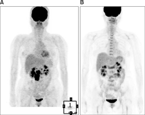
Fig. 1 (A) The [18F]FDG PET/CT showed multiple conglomerated hypermetabolic lymph nodes encasing the inferior vena cava and abdominal aorta in the retrocaval, portocaval and paraaortic spaces. (B) The preoperative [18F]FDG PET/CT revealed that there were no hypermetabolic lesions in the remnant lymph nodes.
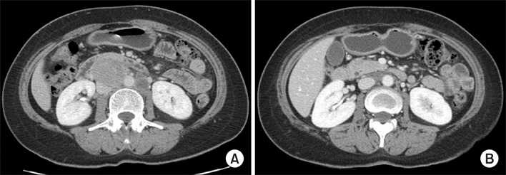
Fig. 2 (A) The initial CT showed huge intraabdominal lymph nodes encasing the inferior vena cava and abdominal aorta. (B) The follow-up CT after 6 cycles of taxane based chemotherapy showed a marked partial response according to the RECIST criteria.

Fig. 3 (A) The laparoscopic biopsy results showed the tumor cells that have pleomorphic nuclei and small cytoplasm (H&E, ×100). (B) The immunohistochemical staining results showed the tumor cells were positive for Wilm's tumor-1. (C) The operative biopsy results showed only extensive necrosis in the remnant lymph nodes without tumor cells (H&E, ×10).
Reference
-
1. Pavlidis N. Cancer of unknown primary: biological and clinical characteristics. Ann Oncol. 2003. 14:Suppl 3. iii11–iii18.2. Greco FA, Hainsworth JD. Introduction: unknown primary cancer. Semin Oncol. 2009. 36:6–7.3. Hainsworth JD, Fizazi K. Treatment for patients with unknown primary cancer and favorable prognostic factors. Semin Oncol. 2009. 36:44–51.4. Fromm GL, Gershenson DM, Silva EG. Papillary serous carcinoma of the peritoneum. Obstet Gynecol. 1990. 75:89–95.5. Dalrymple JC, Bannatyne P, Russell P, Solomon HJ, Tattersall MH, Atkinson K, et al. Extraovarian peritoneal serous papillary carcinoma: a clinicopathologic study of 31 cases. Cancer. 1989. 64:110–115.6. Dennis JL, Hvidsten TR, Wit EC, Komorowski J, Bell AK, Downie I, et al. Markers of adenocarcinoma characteristic of the site of origin: development of a diagnostic algorithm. Clin Cancer Res. 2005. 11:3766–3772.7. Harry VN, Semple SI, Parkin DE, Gilbert FJ. Use of new imaging techniques to predict tumour response to therapy. Lancet Oncol. 2010. 11:92–102.8. Pickles MD, Gibbs P, Lowry M, Turnbull LW. Diffusion changes precede size reduction in neoadjuvant treatment of breast cancer. Magn Reson Imaging. 2006. 24:843–847.
- Full Text Links
-
- Actions
-
Cited
- CITED
-
- Close
- Share
-
- Similar articles
-
- Use of 18F-FDG PET/CT in Second Primary Cancer
- Dual Time Point 18F-FDG PET/CT Imaging Identifies Bilateral Renal Tuberculosis in an Immunocompromised Patient with an Unknown Primary Malignancy
- Late Port Site Metastasis from Occult Gall Bladder Carcinoma After Laparoscopic Cholecystectomy for Cholelithiasis: The Role of 18F-FDG PET/CT
- Lung Adenocarcinoma Staged as an Unknown Primary Presenting with Symptomatic Colon Metastases: Staging by 18F-FDG PET/CT
- Nodular Fasciitis Mimicking Malignant Tumor on 18F-FDG PET/CT
