Dual Time Point 18F-FDG PET/CT Imaging Identifies Bilateral Renal Tuberculosis in an Immunocompromised Patient with an Unknown Primary Malignancy
- Affiliations
-
- 1Department of Nuclear Medicine & PET/CT, Amrita Institute of Medical sciences, Amrita Vishwavidyapeetham University, Cochin, India. padmas@aims.amrita.edu
- KMID: 2068996
- DOI: http://doi.org/10.3947/ic.2015.47.2.117
Abstract
- 18F-FDG PET/CT imaging is an established imaging modality for cancer staging and response assessment. Its role in identifying infective and inflammatory pathologies from malignancy is debated. Dual time - point imaging is a refined technique used to overcome this interpretational dilemma. We present a 59 year old male with an unknown primary malignancy who was referred for a 18F-FDG PET/CT imaging. Images revealed primary lung malignancy with co existing bilateral renal tuberculosis which otherwise would have gone amiss or would have been considered as metastases.
Keyword
MeSH Terms
Figure
-
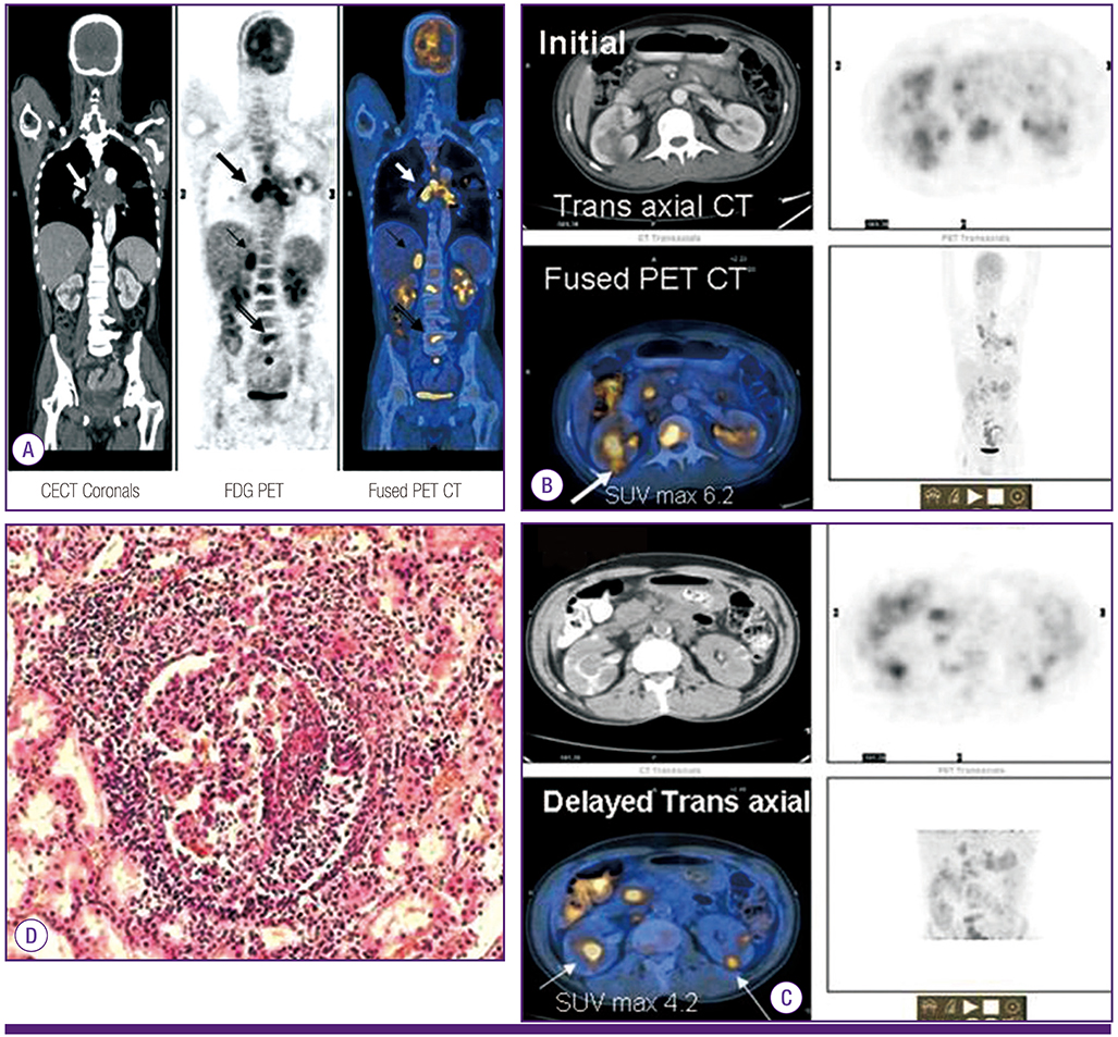
Figure 1 (A) 18F FDG PET/CT imaging in coronal sections showing thick walled irregularly marginated cavitatory lesion in left upper lobe with surrounding lymphangitic spread (SUV Max 3.3) suggestive of primary lung malignancy. Patient also had mediastinal lymphadenopathy (arrows), right adrenal and multiple dorsolumbar vertebral metastatic deposits. (B) Transaxial fused initial PET/CT images further revealed bilateral FDG avid renal lesions (SUV Max of right renal lesion 6.2) (arrow). (C) Delayed transaxial (dual time - point) images showing partial FDG clearance in bilateral renal lesions, marked with arrows (SUV Max 4.2 of right renal lesion). (D) Histology showed extensive caseous necrosis, with occasional granulomas composed of epithelioid cells and Langhans giant cells with surrounding lymphocytes. CECT, contrast-enhanced CT; FDG, flurodeoxyglucose; PET, positron emission tomography; SUV, standardized uptake value.
Reference
-
1. Zhuang H, Pourdehnad M, Lambright ES, Yamamoto AJ, Lanuti M, Li P, Mozley PD, Rossman MD, Albelda SM, Alavi A. Dual time point 18F-FDG PET imaging for differentiating malignant from inflammatory processes. J Nucl Med. 2001; 42:1412–1417.2. Kubota K, Itoh M, Ozaki K, Ono S, Tashiro M, Yamaguchi K, Akaizawa T, Yamada K, Fukuda H. Advantage of delayed whole-body FDG-PET imaging for tumour detection. Eur J Nucl Med. 2001; 28:696–703.
Article
- Full Text Links
-
- Actions
-
Cited
- CITED
-
- Close
- Share
-
- Similar articles
-
- Incidental Bilateral Renal Oncocytoma in a Patient with Metastatic Carcinoma of Unknown Primary: a Pitfall on 18F-FDG PET/CT
- Use of 18F-FDG PET/CT in Second Primary Cancer
- The Clinical Role of Dual-Time-Point 18F-FDG PET/CT in Differential Diagnosis of the Thyroid Incidentaloma
- Bilateral Tubo-Ovarian Abscess Mimics Ovarian Cancer on MRI and 18F-FDG PET/CT
- Lung Adenocarcinoma Staged as an Unknown Primary Presenting with Symptomatic Colon Metastases: Staging by 18F-FDG PET/CT
