J Cardiovasc Ultrasound.
2012 Mar;20(1):57-59. 10.4250/jcu.2012.20.1.57.
Primary Malignant Pericardial Mesothelioma Presenting as Acute Pericarditis
- Affiliations
-
- 1Department of Internal Medicine, Seoul National University College of Medicine, Seoul National University Hospital, Seoul, Korea.
- 2Department of Internal Medicine, Seoul National University College of Medicine, Cardiovascular Center, Seoul National University Bundang Hospital, Seongnam, Korea. cardioch@snu.ac.kr
- KMID: 2177343
- DOI: http://doi.org/10.4250/jcu.2012.20.1.57
Abstract
- We report on a 21-year-old man with fever, dyspnea, and pleuritic chest pain. An electrocardiography showed ST elevation in multiple lead and thoracic echocardiography revealed moderate pericardial effusion. He was initially diagnosed with acute pericarditis, and treated with nonsteroidal anti-inflammatory drugs and colchicines with clinical and laboratory improvement. After 1 month of medication, his symptoms recurred. An echocardiography showed constrictive physiology and the patient was treated with steroid on the top of current medication. The patient had been well for 7 months until dyspnea and edema developed, when an echocardiography showed marked increased pericardial thickness and constriction. Pericardial biopsy was performed and primary malignant pericardial mesothelioma was diagnosed. Malignancy should be considered in the differential diagnosis of recurrent pericarditis.
MeSH Terms
Figure
-

Fig. 1 Pericardial effusion on initial echocardiographic evaluation.
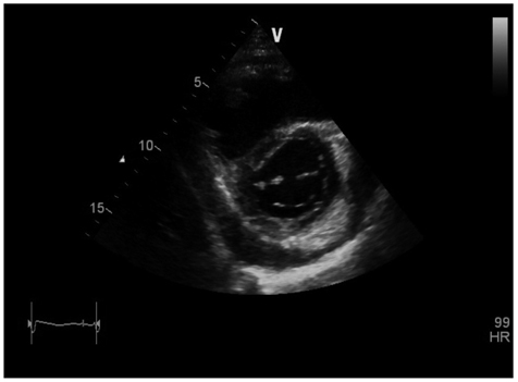
Fig. 2 Moderate amount of pericardial effusion with adhesion after 1 month of treatment with nonsteroidal anti-inflammatory drugs and colchicines.
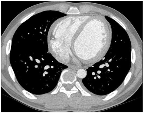
Fig. 3 Improved pericardial effusion with normal pericardial thickness after 4 days of systemic steroid treatment.
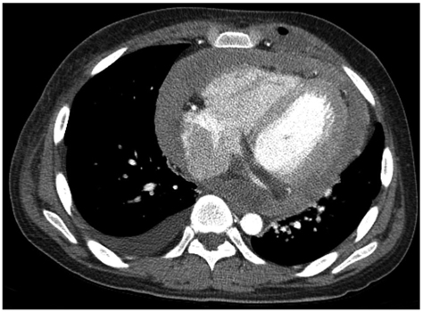
Fig. 4 Diffuse increased pericardial thickening with pericardial enhancement.
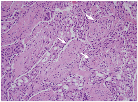
Fig. 5 Atypical mesothelial proliferation with papillary growth configuration and nuclear pleomorphism (H&E stain, ×200; scale bar: 40 µm). White arrows: papillary growth configuration.
Reference
-
1. Patel J, Sheppard MN. Primary malignant mesothelioma of the pericardium. Cardiovasc Pathol. 2011. 20:107–109.
Article2. Thomason R, Schlegel W, Lucca M, Cummings S, Lee S. Primary malignant mesothelioma of the pericardium. Case report and literature review. Tex Heart Inst J. 1994. 21:170–174.3. Khandaker MH, Espinosa RE, Nishimura RA, Sinak LJ, Hayes SN, Melduni RM, Oh JK. Pericardial disease: diagnosis and management. Mayo Clin Proc. 2010. 85:572–593.
Article4. Gössinger HD, Siostrzonek P, Zangeneh M, Neuhold A, Herold C, Schmoliner R, Laczkovics A, Tscholakoff D, Mösslacher H. Magnetic resonance imaging findings in a patient with pericardial mesothelioma. Am Heart J. 1988. 115:1321–1322.
Article5. Kaminaga T, Takeshita T, Kimura I. Role of magnetic resonance imaging for evaluation of tumors in the cardiac region. Eur Radiol. 2003. 13:Suppl 6. L1–L10.
Article6. Papi M, Genestreti G, Tassinari D, Lorenzini P, Serra S, Ricci M, Pasquini E, Nicolini M, Pasini G, Tamburini E, Fattori PP, Ravaioli A. Malignant pericardial mesothelioma. Report of two cases, review of the literature and differential diagnosis. Tumori. 2005. 91:276–279.
Article
- Full Text Links
-
- Actions
-
Cited
- CITED
-
- Close
- Share
-
- Similar articles
-
- A Case of Malignant Pericardial Mesothelioma With Constrictive Pericarditis Physiology Misdiagnosed as Pericardial Metastatic Cancer
- Malignant Pericardial Mesothelioma Misdiagnosed as Constrictive Pericarditis
- A Case of Primary Pericardial Malignant Mesothelioma
- A case of malignant pericardial mesothelioma misdiagnosed as tuberculosis pericarditis
- Surgical Experience of Pericardial Mesothelioma: 2 Cases
