J Korean Acad Conserv Dent.
2006 Jan;31(1):20-29. 10.5395/JKACD.2006.31.1.020.
The effect of restorative materials on the stress distribution of class V composite resin restorations: a 3D finite element investigation
- Affiliations
-
- 1Department of Conservative Dentistry, School of Dentistry, Pusan National University, Korea. jeongkil@pusan.ac.kr
- KMID: 2175777
- DOI: http://doi.org/10.5395/JKACD.2006.31.1.020
Abstract
- The purpose of this study was to analyze the stress distribution aspect of unrestored and restored combined shape (wedge shape occlusally and saucer shape gingivally) class V cavity, which found frequently in clinical cases. A maxillary second premolar restored with a combined shape class V composite restorations were modeled using the three dimensional finite element method. Static occlusal load of 170 N was applied on lingual incline of buccal cusp at the angle of 45degrees with the longitudinal axis of the tooth. And three dimensional finite element analysis was taken by ANSYS (Version 6.0, Swanson Analysis System Co., Houston, U.S.A) program which represent the stress distribution on unrestored and restored cavity wall and margin. The conclusions were as follows. 1. Compared to the unrestored cavity, Von Mises stress at the cementoenamel junction and line angle of the cavity base were reduced and in restored cavity. 2. Von Mises stress at the occlusal and cervical cavity margin and wall were increased in restored cavity in comparison with the unrestored cavity. 3. In the hybrid and hybrid/flowable composite resin restoration, Von Mises stress at the cementoenamel junction and line angle of the cavity base were reduced more than in the flowable restoration. 4. In the hybrid and hybrid/flowable composite resin restoration, Von Mises stress at the occlusal and cervical cavity margin and wall were increased more than in the flowable restoration.
Keyword
Figure
-
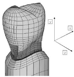
Figure 1 The 3-D finite element mesh model.
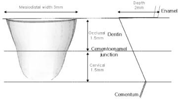
Figure 2 Cavity Design.
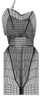
Figure 3 Applied force on the tooth.
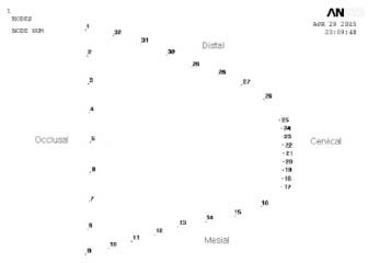
Figure 4 Von Mises stress node number of cavity outline.

Figure 5 Von Mises stress node number of cavity wall.
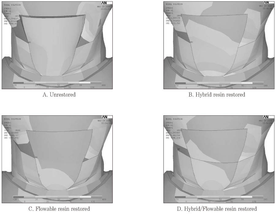
Figure 6 Stress distribution on cavity margins and restorations.
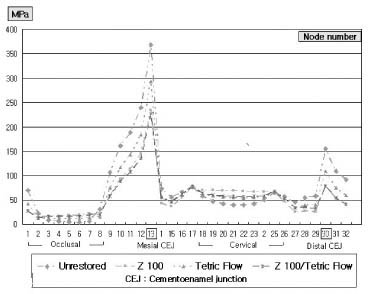
Figure 7 Stress distribution on cavity margins (before and after restoration).

Figure 8 Stress distribution on cavity walls.
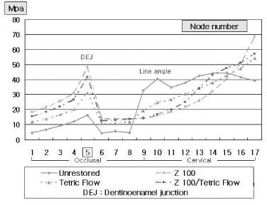
Figure 9 Stress distribution on cavity wall (before and after filling).
Cited by 1 articles
-
Comparison of marginal microleakage between low and high flowable resins in class V cavity
Sang-Bae Bae, Young-Gon Cho, Myeong-Seon Lee
J Korean Acad Conserv Dent. 2009;34(6):477-483. doi: 10.5395/JKACD.2009.34.6.477.
Reference
-
1. Grippo JO, Simring M, Schreiner S. Attrition, abrasion, corrosion and abfraction revisited: A new perspective on tooth surface lesions. J Am Dent Assoc. 2004. 135(8):1109–1118.2. Gallien GS, Kaplan I, Owens BM. A review of noncarious dental cervical lesions. Compendium. 1994. 15(11):1366–1374.3. Owens BM, Gallien GS. Noncarious dental "abfraction" lesion in aging population. Compend Contin Educ Dent. 1995. 16:552–561.4. Cho IS, Park JI, Kwon HC. The microleakage of light-cured glass lonomer restorative materials in class v cavities. J Korean Acad Conserv Dent. 1998. 23(1):304–315.5. Minakuchi S, Munoz CA, Jessop N. Effect of flexural load cycling on microleakge of extended root caries restorations. Oper Dent. 2005. 30(2):234–238.6. Yazici Ar, Celik C, Ozgunaltay G. Microleakage of different resin composite types. Quintessence Int. 2004. 35(10):790–794.7. Park JK, Hur B, Lee HJ. The effect of cavity configuration on marginal leakage of class 5 restoration. J Korean Acad Conserv Dent. 2001. 26(2):162–170.8. Jang HJ, Lee HJ, Hur B. Comparison of marginal leakage of wedge-shaped class v cavity according to restorative materials. J Korean Acad Conserv Dent. 2000. 25(1):56–62.9. Perdigao J, Lambrechts P, Van Meerbeek B, Braem M, Tildiz E, Yucel T, Vanherle G. The interaction of adhesive systems with human dentin. Am J Dent. 1996. 9(4):167–173.10. Braga RR, Cesar PF, Gonzaga CC. Tensile bond strength of filled and unfilled adhesives to dentin. Am J Dent. 2000. 13(2):73–76.11. Lee HE, Lin CL, Wang CH, Chen Ch, Chang CH. Stresses at the cervical lesion of maxillary premolar - a finite element investigation. J Dent. 2002. 30(7-8):283–290.
Article12. Rees JS, Hammadeh M, Jagger DC. Abfraction lesion formation in maxillary incisors, canines and premolars: a finite element study. Eur J Oral Sci. 2003. 111(2):149–154.
Article13. Son YH, Cho BH, Um CM. Finite element stress analysis of class v composite resin restoration subjected to cavity forms and placement methods. J Korean Acad Conserv Dent. 2000. 25(1):91–108.14. Litonjua LA, Bush PJ, Andreana S, Tobias TS, Cohen RE. Effects of occlusal load on cervical lesions. J Oral Rehabil. 2004. 31(3):225–232.
Article15. Rees JS. The effect of variation in occlusal loading on the development of abfraction lesions: a finite element study. J Oral Rehabil. 2002. 29(2):188–193.
Article16. Yaman SD, Sahin M, Aydin C. Finite element analysis of strength characteristics of various resin based restorative materials in Class V cavities. J Oral Rehabil. 2003. 30(6):630–641.
Article17. Palamara D, Palamara JE, Tyas MJ, Messer HH. Strain patterns in cervical enamel of teeth subjected to occlusal loading. Dent Mater. 2000. 16(6):412–419.
Article18. Fruits TJ, VanBrunt CL, Khajotia SS, Duncanson MG Jr. Effect of cyclical lateral forces on microleakage in cervical resin composite restorations. Quintessence Int. 2002. 33(3):205–212.19. Leinfelder KF. Restoration of abfracted lesions. Compendium. 1994. 15(11):1396–1400.20. Heymann HO, Sturdevant JR, Bayne S, Wilder AD, Sluder TB, Brunson WD. Examining tooth flexure effects on cervical restorations: a two-year clinical study. J Am Dent Assoc. 1991. 122(5):41–47.
Article21. Kemp-Scholte CM, Davidson CL. Marginal integrity related to bond strength and strain capacity of composite resin restorative systems. J Prosthet Dent. 1990. 64(6):658–664.
Article22. Bayne SC, Thompson JY, Swift EJ Jr, Stamatiades P, Wilkerson M. A characterization of first-generation flowable composite. J Am Dent Assoc. 1998. 129(5):567–577.23. Gomec Y, Dorter C, Dabanoglu A, Koray F. Effect of resin-based material combination on the compressive and the flexural strength. J Oral Rehabil. 2005. 32(2):122–127.
Article24. Katona TR, Winkler MM. Stress analysis of a bulk-filled Class V light-cured composite resotration. J Dent Res. 1994. 73(8):1470–1477.
Article25. Geramy A, Sharafoddin F. Abfraction: 3D analysis by means of the finite element method. Quintessence Int. 2003. 34(7):526–533.26. Rees JS, OIDougherty D, Pullin R. The stress reducing capacity of unfilled resin in a Class V cavity. J Oral Rehabil. 1999. 26(5):422–427.
Article27. Kim IC. A Study of the Gnathodynamometer. Korean Med J. 1963. 8(11):28. Gibbs CH, Mahan PE, Lundeen HC, Brehnan K, Walsh EK, Sinkewiz SL, Ginsberg SB. Occlusal forces during chewing-Influences of biting strength and food consistency. J Prosthet Dent. 1981. 46(5):561–567.
Article29. Cavel WT, Kelsey WP, Blankenau RJ. An in vivo study of cuspal fracture. J Prosthet Dent. 1985. 53(1):38–42.30. Yettram AL, Wright KW, Pickard HM. Finite element stress analysis of the crowns of normal and restored teeth. J Dent Res. 1976. 55(6):1004–1011.
Article31. Rees JS. A review of the biomechanics of abfraction. Eur J Prosthodont Restor Dent. 2000. 8(4):139–144.32. Kuroe T, Itoh H, Caputo AA, Konuma M. Biomechanics of cervical tooth structure lesions and their restoration. Quintessence Int. 2000. 31(4):267–274.33. Rees JS, Hammadeh M. Undermining of enamel as a mechanism of abfraction lesion formation: a finite element study. Eur J Oral Sci. 2004. 112(4):347–352.
Article34. Verdonschot N, Fennis WM, Kuijs RH, Stolk J, Kreulen CM, Creugers NH. Generation of 3-D finite element models of restored human teeth using micro-CT techniques. Int J Prosthodont. 2001. 14(4):310–315.35. Kuroe T, Caputo AA, Ohata N, Itoh H. Biomechanical effects of cervical lesions and restoration on periodontally compromised teeth. Quintessence Int. 2001. 32(2):111–118.
- Full Text Links
-
- Actions
-
Cited
- CITED
-
- Close
- Share
-
- Similar articles
-
- Stress distribution of Class V composite resin restorations: A three-dimensional finite element study
- The effect of the amount of interdental spacing on the stress distribution in maxillary central incisors restored with porcelain laminate veneer and composite resin: A 3D-finite element analysis
- The influence of occlusal loads on stress distribution of cervical composite resin restorations: A three-dimensional finite element study
- The influence of combining composite resins with different elastic modulus on the stress distribution of Class V restoration: a three-dimensional finite element study
- Effect of restoration type on the stress distribution of endodontically treated maxillary premolars; Three-dimensional finite element study
