The influence of combining composite resins with different elastic modulus on the stress distribution of Class V restoration: a three-dimensional finite element study
- Affiliations
-
- 1Department of Conservative Dentistry, School of Dentistry, Pusan National University, Busan, Korea.
- 2Department of Conservative Dentistry, School of Dentistry, Kyungpook National University, Daegu, Korea. skykim@knu.ac.kr
- KMID: 2175981
- DOI: http://doi.org/10.5395/JKACD.2008.33.3.184
Abstract
- This study was to investigate the influence of combining composite resins with different elastic modulus, and occlusal loading condition on the stress distribution of restored notch-shaped non-carious cervical lesion using 3D finite element (FE) analysis.
The extracted maxillary second premolar was scanned serially with Micro-CT. The 3D images were processed by 3D-DOCTOR. ANSYS was used to mesh and analyze 3D FE model. A notch-shaped cavity was modeled and filled with hybrid, flowable resin or a combination of both. After restoration, a static load of 500N was applied in a point-load condition at buccal cusp and palatal cusp. The stress data were analyzed using analysis of principal stress.
Results
showed that combining method such that apex was restored by material with high elastic modulus and the occlusal and cervical cavosurface margin by small amount of material with low elastic modulus was the most profitable method in the view of tensile stress that was considered as the dominant factor jeopardizing the restoration durability and promoting the lesion progression.
Keyword
Figure
-

Figure 1 3 dimensional finite element model of notch shaped cavity.

Figure 2 Simulated restorations of notch-shaped NCCL (Upper left; Z100-dark brown, Upper right; Tetric Flow-light brown, Lower left; Combination 1, Lower right; Combination 2).
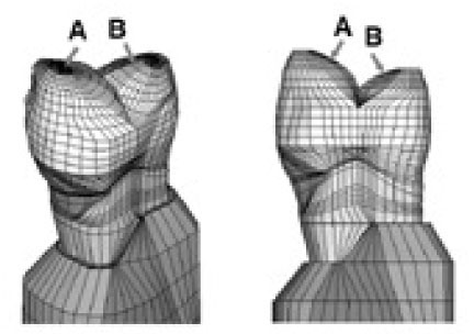
Figure 3 Schematic diagram of loading points (A; Perpendicular load on the upper third of the palatal slope of the buccal cusp, B; perpendicular load on the upper third of the buccal slope of the palatal cusp).
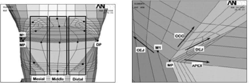
Figure 4 Node selection (left) and magnified aspects (right) of mesial corner of notch-shaped lesion. Along the cavity 4 lines, each 4 nodes were selected at the 3 areas (MP: Mesial point angle, DP: Distal point angle, M1: Nodes near mesial point angle, CEJ: Cemento-enamel junction, Occ: Occlusal cavosurface margin, DEJ: Dentino-enamel junction, Apex: Lesion apex).
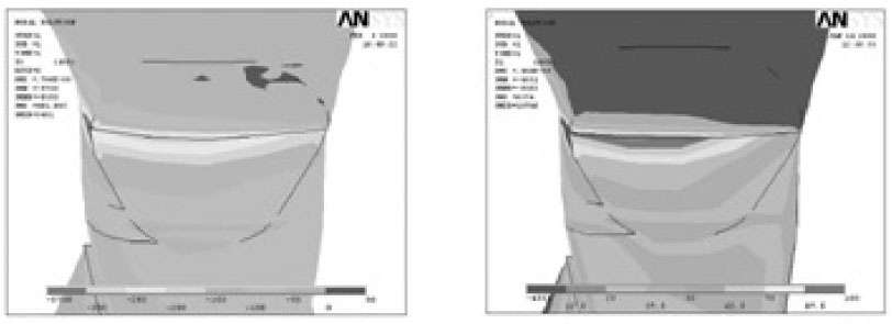
Figure 5 Principal stress distribution of notch-shaped cavity before restoration (Left; Minimum principal stress-Compressive stress, Right; Maximum principal stress-Tensile stress. Upper and lower view's scales were different each other).
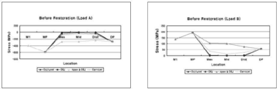
Figure 6 The compressive and tensile stress distribution on occlusal cavosurface margin (Occlusal), DEJ, lesion apex and CEJ (Apex & CEJ), cervical cavosurface margin (Cervical) under Load B (MP: Mesial point angle, DP: Distal point angle, Mes: Mesial node, Mid: Middle node, Dist: Distal node).
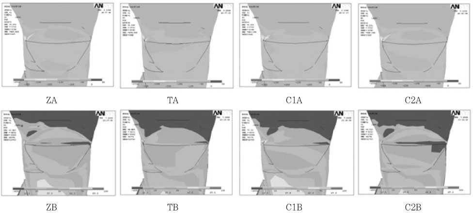
Figure 7 The Principal stress distribution under Load A and B.
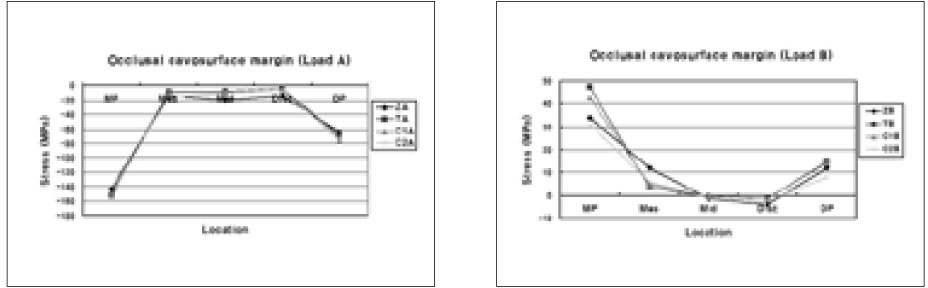
Figure 8 The compressive and tensile stress distribution on occlusal cavosurface margin after restoration under Load B (MP: Mesial point angle, DP: Distal point angle, Mes: Mesial node, Mid: Middle node, Dist: Distal node).
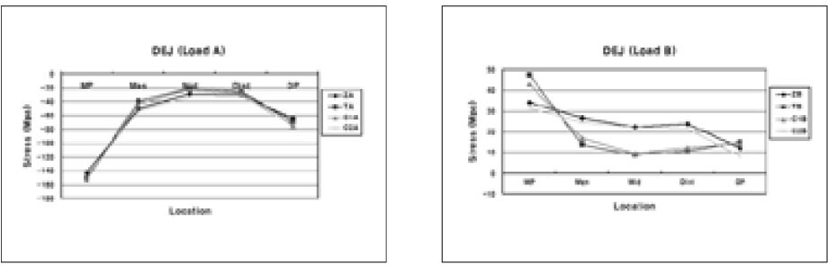
Figure 9 The compressive and tensile stress distribution on occlusal DEJ after restoration under Load B (MP: Mesial point angle, DP: Distal point angle, Mes: Mesial node, Mid: Middle node, Dist: Distal node).

Figure 10 The compressive and tensile stress distribution on lesion apex and CEJ after restoration under Load B (MP: Mesial point angle, DP: Distal point angle, Mes: Mesial node, Mid: Middle node, Dist: Distal node).
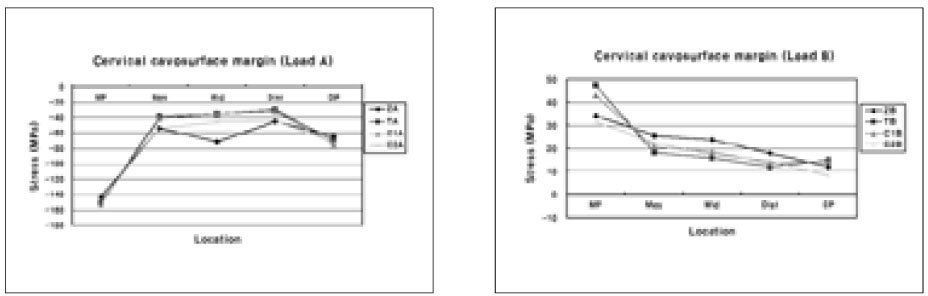
Figure 11 The compressive and tensile stress distribution on cervical cavosurface margin after restoration under Load B (MP: Mesial point angle, DP: Distal point angle, Mes: Mesial node, Mid: Middle node, Dist: Distal node).
Cited by 4 articles
-
Stress analysis of maxillary premolars with composite resin restoration of notch-shaped class V cavity and access cavity; Three-dimensional finite element study
Seon-Hwa Lee, Hyeon-Cheol Kim, Bock Hur, Kwang-Hoon Kim, Kwon Son, Jeong-Kil Park
J Korean Acad Conserv Dent. 2008;33(6):570-579. doi: 10.5395/JKACD.2008.33.6.570.Finite element analysis of maxillary central incisors restored with various post-and-core applications
MinSeock Seo, WonJun Shon, WooCheol Lee, Hyun-Mi Yoo, Byeong-Hoon Cho, Seung-Ho Baek
J Korean Acad Conserv Dent. 2009;34(4):324-332. doi: 10.5395/JKACD.2009.34.4.324.Stress distribution of endodontically treated maxillary second premolars restored with different methods: Three-dimensional finite element analysis
Dong-Yeol Lim, Hyeon-Cheol Kim, Bock Hur, Kwang-Hoon Kim, Kwon Son, Jeong-Kil Park
J Korean Acad Conserv Dent. 2009;34(1):69-79. doi: 10.5395/JKACD.2009.34.1.069.Effect of restoration type on the stress distribution of endodontically treated maxillary premolars; Three-dimensional finite element study
Heun-Sook Jung, Hyeon-Cheol Kim, Bock Hur, Kwang-Hoon Kim, Kwon Son, Jeong-Kil Park
J Korean Acad Conserv Dent. 2009;34(1):8-19. doi: 10.5395/JKACD.2009.34.1.008.
Reference
-
1. Levitch LC, Bader JD, Shugars DA, Heymann HO. Non-carious cervical lesion. J Dent. 1994. 22:195–207.2. Litonjua LA, Bush PJ, Andreana S, Tobias TS. Effects of occlusal load on cervical lesion. J Oral Rehabil. 2004. 31:225–232.3. Telles D, Pegoraro LF, Pereira JC. Prevalence of noncarions cervical lesions and their relation to occlusal aspects: a clinical study. J Esthet Dent. 2000. 12:10–15.
Article4. Palamara D, Palamara JE, Tyas MJ, Messer HH. Strain patterns in cervical enamel of teeth subjected to occlusal loading. Dent Mater. 2000. 16:412–419.
Article5. Lee WC, Eakle WS. Possible role of tensile stress in etiology of cervical erosive lesions of teeth. J Prosthet Dent. 1984. 52:374–380.
Article6. Grippo JO. Abfractions: A new classification of hard tissue lesions of tooth. J Esthet Dent. 1991. 3:14–19.7. Kuroe T, Caputo AA, Ohata N, Itoh H. Biomechanical effects of cervical lesions and restoration on periodontally compromised teeth. Quintessence Int. 2001. 32:111–118.8. King PA. Adhesive techniques. Br Dent J. 1999. 186:321–326.
Article9. Osborne-Smith KL, Burke FJ, Wilson NH. The aetiology of the non-carious cervical lesion. Int Dent J. 1999. 49:139–143.
Article10. Vandewalle KS, Vigil G. Guidelines for the restoration of class V lesions. Gen Dent. 1997. 45:254–260.11. Leinfelder KF. Restoration of abfracted lesions. Compendium. 1994. 15:1396–1400.12. Heymann HO, Sturdevant JR, Bayne S, Wilder AD, Sluder TB, Brunson WD. Examining tooth flexure effects on cervical restorations: a two-year clinical study. J Am Dent Assoc. 1991. 122:41–47.
Article13. Loguercio AD, Zago C, Leal K, Ribeiro NR, Reis A. One-year clinical evaluation of flowable resin liner associated with a microhybrid resin in noncarious cervical lesions. Clin Oral Investig. 2005. 9:18–20.
Article14. Park JK, Hur B, Kim SK. Stress distribution of class V composite resin restorations: A three-dimensional finite element study. J Korean Acad Conserv Dent. 2008. 33:28–38.
Article15. Lindehe J, Karring T. Textbook of Clinical Periodontology. 1989. 2nd edition. Copenhagen: Munksgaard;19–69.16. Schroeder HE, Page RC. Periodontal Diseases. 1990. 2nd edition. Philadelphia: Lea & Fabiger;3–52.17. Rubin C, Krishnamurthy N, Capilouto E, Yi H. Stress analysis of the human tooth using a three-dimensional finite element model. J Dent Res. 1983. 62:82–86.18. Katona TR, Winkler MM. Stress analysis of a bulk-filled class V light-cured composite restoration. J Dent Res. 1994. 73:1470–1477.
Article19. Geramy A, Sharafoddin F. Abfraction: 3D analysis by means of the finite element method. Quintessence Int. 2003. 34:526–533.20. Le SY, Chiang HC, Huang HM, Shih YH, Chen HC, Dong DR, Lin CT. Thermo-debonding mechanisms in dentin bonding systems using finite element analysis. Biomaterials. 2001. 22:113–123.
Article21. Kleverlaan CJ, Feilzer AJ. Polymerization shrinkage and contraction stress of dental resin composite. Dent Mater. 2005. 21:1150–1157.
Article22. Litonjua LA, Andreana S, Patra AK, Cohen RE. An assessment of stress analyses in the theory of abfraction. Biomed Mater Eng. 2004. 14:311–321.23. Dejak B, Mlotkowski A, Romanowicz M. Finite element analysis of mechanism of cervical lesion formation in simulated molars during mastication and parafunction. J Prosthet Dent. 2005. 94:520–529.
Article24. Kuroe T, Itoh H, Caputo AA, Konuma M. Biomechanics of cervical tooth structure lesions and their restoration. Quintessence Int. 2000. 31:267–274.25. Lee WC, Eakle WS. Stress-induced cervical lesions: review of advances in the past 10 years. J Prosthet Dent. 1996. 75:487–494.
Article26. Neo J, Chew C, Yap A, Sidhu S. Clinical evaluation of tooth-colored materials in cervical lesions. Am J Dent. 1996. 9:15–17.27. Kemp-Scholte CM, Davidson CL. Marginal integrity related to bond strength and strain capacity of composite resin restorative systems. J Prosthet Dent. 1990. 64:658–664.
Article28. Rees JS, Hammadeh M. Undermining of enamel as a mechanism of abfraction lesion formation: a finite element study. Eur J Oral Sci. 2004. 112:347–352.
Article29. Yaman SD, Sahin M, Aydin C. Finite element analysis of strength characteristics of various resin based restorative materials in class V cavities. J Oral Rehabil. 2003. 30:630–641.
Article30. Nakayama WT, Hall DR, Grenoble DE, Katz JL. Elastic properties of dental resin restorative materials. J Dent Res. 1974. 53:1121–1126.
Article31. Willems G, Lambrechts P, Braem M, Celis JP, Vanherle G. A classification of dental composites according to their morphological and mechanical characteristics. Dent Mater. 1992. 8:310–319.
Article32. Heymann HO, Sturdevant JR, Bayne S, Wilder AD, Sluder TB, Brunson WD. Examining tooth flexure effects on cervical restorations: a two-year clinical study. J Am Dent Assoc. 1991. 122:41–47.
Article33. Braga RR, Hilton TJ, Ferracane JL. Contraction stress of flowable composite materials and their efficacy as stress-relieving layers. J Am Dent Assoc. 2003. 134:721–728.
Article
- Full Text Links
-
- Actions
-
Cited
- CITED
-
- Close
- Share
-
- Similar articles
-
- The effects of dentin bonding agent thickness on stress distribution of composite-tooth interface : Finite element method
- Stress distribution of Class V composite resin restorations: A three-dimensional finite element study
- The influence of occlusal loads on stress distribution of cervical composite resin restorations: A three-dimensional finite element study
- Stress distribution of endodontically treated maxillary second premolars restored with different methods: Three-dimensional finite element analysis
- Influence of various properties of post and core on the stress distribution in endodontically treated tooth
